- Clone
- 105 (See other available formats)
- Regulatory Status
- RUO
- Other Names
- F12A2B5, 105
- Isotype
- Mouse IgM, κ

-

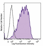
Insulinoma cell line, Beta-TC-6, was stained with purified A2B5 (clone 105, filled histogram) or mouse IgM, κ isotype control (open histogram) followed by anti-mouse IgM PE.
| Cat # | Size | Price | Quantity Check Availability | ||
|---|---|---|---|---|---|
| 150702 | 100 µg | $206.00 | |||
The 105/A2B5 antibody recognizes a membrane protein that is expressed by chicken retinal cells, neuronal cells, ganglia, and spinal cord. In combination with other markers, this antibody can be used to differentiate between type 1 and type 2 astrocytes. The antibody recognizes polysialogangliosides and can be used to monitor glial cell development. A2B5 antigens are expressed by a variety of tumors including glioblastomas. Tumors expressing A2B5 antigen exhibit higher recurrence. The A2B5 antibody can also be used to identify progenitor cells.
Product Details
- Verified Reactivity
- Mouse, Human
- Antibody Type
- Monoclonal
- Host Species
- Mouse
- Immunogen
- Embryonic chicken retinal cells.
- Formulation
- Phosphate-buffered solution, pH 7.2, containing 0.09% sodium azide.
- Preparation
- The antibody was purified by affinity chromatography.
- Concentration
- 0.5 mg/ml
- Storage & Handling
- The antibody solution should be stored undiluted between 2°C and 8°C.
- Application
-
FC - Quality tested
IHC-F - Reported in the literature, not verified in house - Recommended Usage
-
Each lot of this antibody is quality control tested by immunofluorescent staining with flow cytometric analysis. For flow cytometric staining, the suggested use of this reagent is ≤ 1.0 µg per million cells in 100 µl volume. It is recommended that the reagent be titrated for optimal performance for each application.
- Application Notes
-
Additional reported applications (for the relevant formats of this clone) include: immunofluorescence staining.1,2
-
Application References
(PubMed link indicates BioLegend citation) -
- Eisenbarth GS, et al. 1979. Proc. Natl. Acad. Sci. 76:4913. (IF)
- Eisenbarth GS, et al. 1982. Proc. Natl. Acad. Sci. 79:5066. (IF)
- Russell TR, et al. 1984. Cytometry 5:539. (FC)
- RRID
-
AB_2566277 (BioLegend Cat. No. 150702)
Antigen Details
- Distribution
-
Developing thymic epithelial cells, oligodendrocyte progenitors, neuroendocrine, embryonic neonatal tissue, astrocytoma, glioma, and squamous cell carcinoma.
- Cell Type
- Epithelial cells, Oligodendrocytes
- Biology Area
- Cell Biology, Immunology, Neuroscience, Neuroscience Cell Markers
- Antigen References
-
1. Baracskay KL, et al. 2007. GLIA 55:1001.
2. Raff MC, et al. 1983. J. Neurosci. 6:1289.
3. Fredman P, et al. 1984. Arch. Biochem. Biophys. 2:661.
4. Xia CL, et al. 2003. Int. J. Oncol. 23:353.
5. Tchoghandjian A, et al. Brain Pathol. 20:211. - Gene ID
- NA
- UniProt
- View information about A2B5 on UniProt.org
Other Formats
View All A2B5 Reagents Request Custom Conjugation| Description | Clone | Applications |
|---|---|---|
| Purified anti-mouse/human A2B5 | 105 | FC,IHC-F |
| Alexa Fluor® 647 anti-mouse/human A2B5 | 105 | FC |
Compare Data Across All Formats
This data display is provided for general comparisons between formats.
Your actual data may vary due to variations in samples, target cells, instruments and their settings, staining conditions, and other factors.
If you need assistance with selecting the best format contact our expert technical support team.
-
Purified anti-mouse/human A2B5

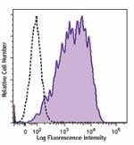
Insulinoma cell line, Beta-TC-6, was stained with purified A... -
Alexa Fluor® 647 anti-mouse/human A2B5

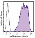
Insulinoma cell line, Beta-TC-6, was stained with A2B5 (clon...
