- Regulatory Status
- RUO
- Other Names
- Tag-It Violet Proliferation Cell Tracking Dye

-

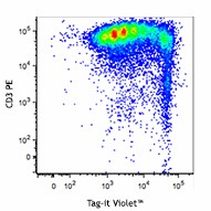
Human peripheral blood mononuclear cells were stained with Tag-it Violet™ Proliferation and Cell Tracking dye, and then stimulated with PHA for four days. On day four, cells were harvested, stained with CD3-PE and the Tag-it Violet™ signal was analyzed by flow cytometry. -

Mouse spleen 72 hours after adoptive transfer of Tag-it Violet™-labeled splenocytes (purple). Nucleated cells are stained using 25 µM DRAQ5™ (red). Image was captured at a 40X magnification.
| Cat # | Size | Price | Quantity Check Availability | ||
|---|---|---|---|---|---|
| 425101 | 1 kit | $265.00 | |||
Tag-it Violet™ Proliferation and Cell Tracking Dye can be used for cell tracking and for proliferation assays. Tag-it Violet™ passively diffuses into the cell where esterases cleave acetoxymethyl esters (AM) on the molecule, Tag-it Violet™ then covalently attaches to intracellular proteins enabling its long-term retention.
Tag-it Violet™ is excited with a fluorescence wavelength of 395 nm, and emits at 455 nm. Cells labeled wtih Tag-it Violet™ can be analyzed by flow cytometry in instruments equipped with a violet laser, or visualized by fluorescence microscopy using a DAPI filter.
Product Details
- Preparation
- The Tag-it Violet™ Proliferation and Cell Tracking Dye is composed of lyophilized Tag-it Violet™ and anhydrous DMSO. For reconstitution, bring the kit to room temperature, add 50 µl of DMSO to one vial of Tag-it Violet™ dye until fully dissolved.
- Storage & Handling
- The Tag-it Violet™ Proliferation and Cell Tracking Dye should be stored at -20°C upon receipt. Do not open vials until needed. Once the DMSO is added to the Tag-it Violet™, use immediately, or store at -20°C in a dry place and protected ifrom light, preferably in a dessicator or in a container with dessicant for no more than one month.
- Application
-
FC - Quality tested
ICC - Verified - Recommended Usage
-
This lot has been tested by flow cytometry for in vitro cell proliferation. It can be used at concentrations ranging from 1 - 20 µM for cell labeling. It is recommended that the reagent be titrated for optimal performance for each cell type, culturing condition, or application.
- Application Notes
-
The molecular weight of Tag-it Violet™ Proliferation and Cell Tracking Dye is 489 kD. The maximum excitation and emission wavelengths of Tag-it Violet™ Proliferation and Cell Tracking Dye are 395 nm and 455 nm, respectively. Each 122.25 µg of Tag-it Violet™ Proliferation and Cell Tracking Dye may be reconstituted with 50 µl of anhydrous DMSO to yield a stock concentration of 5 mM.
Materials Provided:
5 vials x 122.25 µg Tag-it Violet™
500 µl anhydrous DMSO
Tag it-Violet™ Labeling Procedure:
1. Prior to reconstitution, spin down the vial of lyophilized reagent in a microcentrofuge to ensure the reagent is at the bottom of the vial.
2. Prepare stock solution by reconstituting 1 vial of lyophilized Tag-it Violet™ dye in 50 µl of anhydrous DMSO to make a 5 mM solution.
3. Prepare a 5 µM working solution by diluting 1 µL of 5 mM Tag-it Violet™ stock solution in 1 mL PBS for every 1 mL of cell suspension (or at an optimal working concentration as determined by titration).
4. Spin down and resuspend cells at 10-100 x 106 cells/mL in the Tag-it Violet™ working solution.
5. Incubate cells for 20 minutes at room temperature or at 37°C, and keep protected from light.
6. Quench the staining by adding five times the original staining volume of cell culture medium containing 10% FBS.
7. Pellet cells and resuspend in pre-warmed cell culture medium.
8. Incubate cells for 10 minutes.
9. After incubation, Tag-it Violet™ labeled cells are ready for downstream applications or analysis. - Product Citations
-
Antigen Details
- Structure
- 489 kD.
- Biology Area
- Cell Biology, Cell Cycle/DNA Replication, Cell Proliferation and Viability, Neuroscience
- Gene ID
- NA
