Zombie UV™ Fixable Viability Kit

Pricing & Availability
Regulatory Status
RUO
Other Names
Fixable Dye, Fixable Viability Dye
Zombie_UV_Viability_Dye_012714
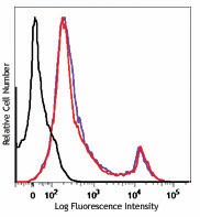
One day old C57BL/6 mouse splenocytes were stained with Zombie UV™ and analyzed before fixation (purple) or after fixation and permeabilization (red). Cells alone, without Zombie UV™ staining, are indicated in black.
  • Zombie_UV_Viability_Dye_012714
    One day old C57BL/6 mouse splenocytes were stained with Zombie UV™ and analyzed before fixation (purple) or after fixation and permeabilization (red). Cells alone, without Zombie UV™ staining, are indicated in black.
See Zombie UV™ spectral data
Cat # Size Price Quantity Check Availability
423107 100 tests $89.00
Check Availability


Need larger quantities of this item?
Request Bulk Quote
423108 500 tests $335.00
Check Availability


Need larger quantities of this item?
Request Bulk Quote
Description

Zombie UV™ is an amine reactive fluorescent dye that is non-permeant to live cells, but permeant to the cells with compromised membranes. Thus, it can be used to assess live vs. dead status of mammalian cells. Zombie UV™ is a polar water soluble dye, providing violet fluorescence, making it suitable for multi-color detection.

Technical data sheet

Product Details

Preparation
Zombie UV™ Fixable Viability kit is composed of lyophilized Zombie UV™ dye and anhydrous DMSO. For reconstitution, bring the kit to room temperature; add 100 µl of DMSO to one vial of Zombie UV™ dye until fully dissolved. 100 tests = 1 vial of Zombie UV™ + DMSO, 500 tests = 5 vials of Zombie UV™ + DMSO.
Storage & Handling
Store kit at -20°C upon receipt. Do not open vials until needed. Once the DMSO is added to the Zombie UV™ dye, use immediately, or store at -20°C in a dry place and protected from light, preferably in a desiccator or in a container with desiccant for no more than one month.
Application

FC - Quality tested

Recommended Usage

Each lot of this product is quality control tested by immunofluorescent staining with flow cytometric analysis.

For flow cytometry, the suggested dilution is 1:100-1:1000 for 1-10 million cells. It is recommended that the reagent be titrated for optimal performance for each application, as optimal dosage varies with cell type.

Excitation Laser
Ultraviolet Laser (355 nm)
Application Notes

Standard Cell Staining Protocol:

  1. Prior to reconstitution, spin down the vial of lyophilized reagent in a microcentrofuge to ensure the reagent is at the bottom of the vial.
  2. For reconstitution, pre-warm the kit to room temperature; add 100 µl of DMSO to one vial of Zombie UV™ dye and mix until fully dissolved
  3. Wash cells with PBS buffer (no Tris buffer and protein free).
  4. Dilute Zombie UV™ dye at 1:100-1000 in PBS. Resuspend 1-10 x 106 cells in diluted 100 µl Zombie UV™ solution. To minimize background staining of live cells, titrate the amount of dye and/or number of cells per 100 µl for optimal performance. Different cell types can have a wide degree of variability in staining based on cell size and degree of cell death.

    Note: Don’t use Tris buffer as a diluent and be sure that the PBS does not contain any other protein like BSA or FBS.
    Note: The amount of dye used can also influence the ability to detect apoptotic as well as live and dead cells.

  5. Incubate the cells at room temperature, in the dark, for 15-30 minutes.
  6. Wash one time with 2 ml BioLegend’s Cell Staining Buffer (Cat. No. 420201) or equivalent buffer containing serum or BSA.
  7. Continue performing antibody staining procedure as desired.
  8. Cells can be fixed with paraformaldehyde or methanol prior to permeabilization or can be analyzed without fixation.

No-wash Sequential Staining Protocol:

  1. Wash cells with PBS buffer (no Tris buffer and protein free).
  2. For reconstitution, pre-warm the kit to room temperature; add 100 µl of DMSO to one vial of Zombie UV™ dye and mix until fully  dissolved
  3. Determine the total µl volume of antibody cocktail previously titrated and optimized for the assay that will be added to each vial/well of cells based on a final volume of 100 µl. Subtract that antibody volume from the 100 µl total staining volume intended for the assay. In the remaining volume, dilute Zombie UV™ dye at 1:100-1000 in PBS as determined by prior optimization at that volume. For example, if you are adding 20 µl of antibody cocktail for a 100 µl total staining volume, use 80 µl of Zombie UV™ solution. Resuspend 1-10 x 106 cells in the appropriate volume of Zombie UV™ solution. Different cell types can have a wide degree of variability in staining based on cell size and degree of cell death.

    Note: Don’t use Tris buffer as a diluent and be sure that the PBS does not contain any other protein like BSA or FBS.
    Note: The amount of dye used can also influence the ability to detect apoptotic as well as live and dead cells.

  4. Incubate for 10-15 minutes at RT, protected from light. Without washing the cells, add the cell surface antibody cocktail and incubate for another 15-20 minutes.
  5. Add 1-2 mL Cell Staining Buffer (Cat. No. 420201) or equivalent buffer containing BSA or serum. Centrifuge to pellet.
  6. Continue with normal fixation and permeabilization procedure. If planning to skip fixation and analyze cells live, complete an additional wash step to minimize any unnecessary background of the live cells.


Notes: If the cell type in use cannot tolerate a protein-free environment, then titrate the Zombie UV™ dye in the presence of the same amount of BSA/serum as will be present in the antibody staining procedure. A higher amount of Zombie UV™ may be required since the BSA/serum will react with and bind up some proportion of the Zombie UV™.

Zombie UV™ dye is excited by the UV laser (350 nm) and has fluorescence emission maximum at 459 nm. If using in a multi-color panel design, filter optimization may be required depending on other fluorophores used. Zombie UV™ dye has similar emission to DAPI.


Application References

(PubMed link indicates BioLegend citation)
  1. Souza-Fonesca-Guimaraes F, et al. 2015. PNAS. 112:2376. PubMed
Product Citations
  1. Arruda DC, et al. 2022. J Control Release. 350:228. PubMed
  2. Michaels YS, et al. 2022. Sci Adv. 8:eabn5522. PubMed
  3. Lutz EA, et al. 2022. Proc Natl Acad Sci U S A. 119:e2205983119. PubMed
  4. Bonadonna M, et al. 2022. Sci Adv. 8:eabq4469. PubMed
  5. Humphries DC, et al. 2023. Front Immunol. 14:1100161. PubMed
  6. Heng Y, et al. 2023. J Transl Med. 21:167. PubMed
  7. Yi X, et al. 2023. Signal Transduct Target Ther. 8:107. PubMed
  8. Gu XW, et al. 2023. Nat Commun. 14:1439. PubMed
  9. Arunachalam PS, et al. 2023. J Clin Invest. 133:. PubMed
  10. Hackerova L, et al. 2023. Front Vet Sci. 10:1116891. PubMed
  11. Kak G, et al. 2023. J Neuroinflammation. 20:114. PubMed
  12. Lin W, et al. 2023. Commun Biol. 6:447. PubMed
  13. Borbet TC, et al. 2023. iScience. 26:106810. PubMed
  14. Wang X, et al. 2023. Stem Cell Rev Rep. . PubMed
  15. Souza-Fonseca-Guimaraes F, et al. 2016. Cell Death Dis. 7:e2302. PubMed
  16. Zgair A, et al. 2017. Sci Rep. . 10.1038/s41598-017-15026-z. PubMed
  17. Argüello RJ, et al. 2020. Cell Metab. 32:1063. PubMed
  18. Reyes RA, et al. 2021. PLoS One. 16:e0261656. PubMed
  19. Rosina M, et al. 2022. Cell Metab. 34:533. PubMed
  20. Tocheva AS, et al. 2020. Curr Protoc Immunol. 130:e103. PubMed
  21. Duraiswamy J, et al. 2021. Cancer Cell. 39:1623. PubMed
  22. Klapholz M, et al. 2022. J Pathol. 257:186. PubMed
  23. Alam A, et al. 2022. Cancer Cell. 40:153. PubMed
  24. Huang SSY, et al. 2022. J Cell Mol Med. 26:1714. PubMed
  25. Atalis A, et al. 2022. J Control Release. 347:476. PubMed
  26. Xu C, et al. 2022. iScience. 25:105123. PubMed
  27. Weng S, et al. 2022. Front Immunol. 13:1025931. PubMed
  28. Tatsumi N, et al. 2022. STAR Protoc. 3:101845. PubMed
  29. Giannou AD, et al. 2023. Immunity. 56:125. PubMed
  30. Mallick S, et al. 2023. Transl Res. :. PubMed
  31. Arimoto KI, et al. 2023. Nat Commun. 14:251. PubMed
  32. Smith LA, et al. 2023. Front Oncol. 12:1031174. PubMed
  33. Denis M, et al. 2023. Front Immunol. 13:1011943. PubMed
  34. Labuz DR, et al. 2023. Elife. 12: . PubMed
  35. Shen JZ, et al. 2022. Mol Cell. 82:1123. PubMed
  36. Zhang H, et al. 2022. J Exp Med. 219:. PubMed
  37. Xu H, et al. 2017. EBioMedicine. 10.1016/j.ebiom.2017.03.003. PubMed
  38. Lu C, et al. 2019. Cancer Immunol Res. 7:414. PubMed
  39. Matsui K, et al. 2015. PLoS One. 10: 0137195. PubMed
  40. Clarke F,et al. 2017. PLoS One. 10.1371/journal.pone.0186625. PubMed
  41. Vaughan HJ, et al. 2021. Mol Ther Oncolytics. 21:377. PubMed
  42. Gruber T, et al. 2020. JCI Insight. 5:00. PubMed
  43. Trimaglio G, et al. 2020. Oncoimmunology. 9:1790125. PubMed
  44. Souza-Fonseca-Guimaraes F, et al. 2015. Proc Natl Acad Sci U S A. 112:2376. PubMed
  45. Rao S, et al. 2017. Cell. 168(3):503-516.e12. PubMed
  46. Godbersen-Palmer C, et al. 2020. J Immunol. 204:2973. PubMed
  47. Chaurasiya S, et al. 2020. Oncoimmunology. 9:1729300. PubMed
  48. Rafiq S, et al. 2018. Nat Biotechnol. 36:847. PubMed
  49. Poggio M, et al. 2019. Cell. 177:414. PubMed
  50. Liu Y, et al. 2022. Nat Commun. 13:2665. PubMed
  51. Leigh N, et al. 2017. The Journal of Immunology. 10.4049/jimmunol.1502181. PubMed
  52. An J,et al. 2017. Sci Rep.. 10.1038/s41598-017-13629-0. PubMed
  53. Sellau J, et al. 2020. Nat Commun. 2.860416667. PubMed
  54. Odeh-Couvertier VY, et al. 2022. Bioeng Transl Med. 7:e10282. PubMed
  55. Vanshylla K, et al. 2022. Cell Host Microbe. 30:69. PubMed
  56. Guo W, et al. 2022. J Immunother Cancer. 10:. PubMed
  57. Reighard SD, et al. 2020. Cell Rep Med. :1. PubMed
  58. Lin J, et al. 2017. Sci Rep. 7:41722. PubMed
  59. Lord JD, et al. 2018. Clin Immunol. 193:24:00. PubMed
  60. Nath PR, et al. 2019. Cancer Immunol Res. 7:1547. PubMed
  61. Fallet B, et al. 2020. Cell Rep. 30:1013. PubMed
  62. Littwitz-Salomon E, Schimmer S, and Dittmer U. 2017. J Virol. 10.1128/JVI.01122-17. PubMed
  63. Zhang X, et al. 2022. Cell Rep. 40:111203. PubMed
  64. Trüb M, et al. 2020. J Immunother Cancer. 8:00. PubMed
  65. Shiao SL, et al. 2021. Cancer Cell. 39:1202. PubMed
  66. Kallert S, et al. 2017. Nature Communications. 10.1038/ncomms15327. PubMed
  67. Moreno-Fernandez ME, et al. 2018. JCI Insight. 3. PubMed
  68. Li D, et al. 2019. J Lipid Res. 60:1503. PubMed
  69. Lerrer S, et al. 2021. iScience. 24:103020. PubMed
  70. Grigoryan L, et al. 2022. NPJ Vaccines. 7:55. PubMed
  71. Gehling K, et al. 2022. Life Sci Alliance. 5:. PubMed
  72. Nath PR, et al. 2022. Oncoimmunology. 11:2111909. PubMed
  73. Kalbasi A, et al. 2020. Sci Transl Med. :12. PubMed
  74. Huang SSY, et al. 2021. Biology. 10(8):. PubMed
  75. Zeng Y, et al. 2019. Oncotarget. 10:4479. PubMed
  76. Fredriksson-Lidman K, et al. 2017. PLoS One. 10.1371/journal.pone.0185509. PubMed
  77. Truong AS, et al. 2021. J Clin Invest. 131:. PubMed
  78. Holokai L, et al. 2020. Cancers (Basel). 12:00. PubMed
  79. Burns JC, et al. 2020. eLife. 9:00. PubMed
  80. O’Neill R, et al. 2017. J Immunol. 10.4049/jimmunol.1700380. PubMed
  81. Harder I, et al. 2022. Cells. 11:. PubMed
  82. McVey JC, et al. 2022. iScience. 25:103847. PubMed
  83. Smith CA, et al. 2019. JCI Insight. 4. PubMed
  84. Den Braanker H, et al. 2021. Front Immunol. 12:768113. PubMed
  85. Ganguly S, et al. 2021. Cell Mol Gastroenterol Hepatol. 12:891. PubMed
  86. Henrich IC, et al. 2021. Cancer Res. 81:2171. PubMed
  87. Johnston S, et al. 2021. Elife. 10: . PubMed
  88. Pasciuto E, et al. 2020. Cell. 182:625. PubMed
  89. Zhang R, et al. 2022. Front Pharmacol. 13:870848. PubMed
  90. Graciotti M, et al. 2020. Vaccines (Basel). 8:00. PubMed
  91. Capuccini B, et al. 2016. Sci Rep. 6:39258. PubMed
  92. Dangaj D, et al. 2019. Cancer Cell. 35:885. PubMed
  93. Lyle C, et al. 2019. Sci Rep. 9:20257. PubMed
  94. Cartwright ANR, et al. 2021. Cancer Immunol Res. 9:470. PubMed
  95. Moreno-Fernandez ME, et al. 2021. STAR Protoc. 2:100937. PubMed
  96. Wu J, et al. 2021. STAR Protoc. 2:101022. PubMed
  97. Georg P, et al. 2022. Cell. 185:493. PubMed
  98. Ireland RE, et al. 2022. Viruses. 14:. PubMed
  99. Comte D, et al. 2016. Proc Natl Acad Sci U S A. 113: 9321 - 9326. PubMed
  100. Cantor DJ et al. 2019. Cell reports. 26(1):108-118 . PubMed
  101. Reuschl AK, et al. 2022. Cell Rep. 39:110650. PubMed
  102. Salehi S, et al. 2017. PLoS One. 10.1371/journal.pone.0163614. PubMed
  103. Nath PR, et al. 2019. Front Immunol. 9:2985. PubMed
  104. Bommareddy PK, et al. 2019. J Biol Methods. 6:2. PubMed
  105. Mempin M, et al. 2021. Cancers (Basel). 13:. PubMed
  106. Wimmers F, et al. 2021. Cell. 184:3915. PubMed
  107. Galván-Peña S, et al. 2021. Proc Natl Acad Sci U S A. 118:. PubMed
  108. Chen YL, et al. 2022. Front Neurosci. 16:876582. PubMed
  109. Prosser A, et al. 2021. STAR Protoc. 2:100810. PubMed
  110. Giles D, et al. 2016. PLoS One. 11: 0149783. PubMed
  111. Lacar B, et al. 2016. Nat Commun. 7: 11022. PubMed
  112. Grabiec A, et al. 2017. J Allergy Clin Immunol. 10.1016/j.jaci.2017.03.024. PubMed
  113. Gardner A, et al. 2022. J Immunother Cancer. 10:. PubMed
  114. Lee A, et al. 2022. Nat Commun. 13:549. PubMed
  115. Zeng Y, et al. 2019. FASEB J. 33:6596. PubMed
  116. Mitchell LA, et al. 2019. Oncotarget. 10:2252. PubMed
  117. Li CY, et al. 2022. Int J Mol Sci. 23:. PubMed
  118. Zhong W, et al. 2022. Front Immunol. 13:1001255. PubMed
  119. Gamradt S, et al. 2021. iScience. 24:103312. PubMed

Antigen Details

Biology Area
Apoptosis/Tumor Suppressors/Cell Death, Cell Biology, Neuroscience
Gene ID
NA
Go To Top Version: 4    Revision Date: 05/30/2019

8999 BioLegend Way, San Diego, CA 92121 www.biolegend.com
Toll-Free Phone: 1-877-Bio-Legend (246-5343) Phone: (858) 768-5800 Fax: (877) 455-9587

Login/Register
Forgot your password? Reset Password
Request an Account