 Login/Register


- Clone
- 9C4 (See other available formats)
- Regulatory Status
- RUO
- Other Names
- Ep-CAM, tumor associated calcium signal transducer 1, epithelial cell surface antigen, epithelial glycoprotein 2, EGP2, adenocarcinoma associated antigen, TROP1
- Isotype
- Mouse IgG2b, κ
- Ave. Rating
- Submit a Review
- Product Citations
- publications

-

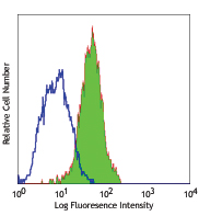
Human colon carcinoma cell line HT29 stained with purified 9C4, followed by anti-mouse IgG FITC -

BT474 breast cancer cells were stained with anti-human CD326 (clone 9C4) using 1:100 dilution, followed by DyLight™ 649 anti-mouse Ig secondary antibody (red) plus DAPI staining for nuclei (blue). Cells were fixed with 4% PFA, permeabilized with 0.1% Triton X-100, blocked with 10% serum, and incubated O/N at 4°C. Images were taken under 20x (filter set: EX 647/10x, Dichroic 665LP, EM 700/70x) at exposure 4s. Data provided by John Nolan and Er Liu, La Jolla Bioengineering Institute.
| Cat # | Size | Price | Quantity Check Availability | Save | ||
|---|---|---|---|---|---|---|
| 324201 | 25 µg | $101 | ||||
| 324202 | 100 µg | $206 | ||||
CD326 is also known as Ep-CAM, tumor associated calcium signal transducer 1, epithelial cell surface antigen, epithelial glycoprotein 2, EGP2, adenocarcinoma associated antigen, and TROP1. CD326 is a type I transmembrane protein containing six disulfide bridges and one THYRO domain. This cell surface glycosylated 40 kD protein is highly expressed in bone marrow, colon, lung, and most normal epithelial cells and is expressed on carcinomas of gastrointestinal origin. Recently, it has been reported that CD326 expression occurs during the early steps of erythrogenesis. CD326 functions as a homotypic calcium-independent cell adhesion molecule and is believed to be involved in carcinogenesis by its ability to induce genes involved in cellular metabolism and proliferation. CD326 antigen is an immunotherapeutic target for the treatment of human carcinomas.
Product DetailsProduct Details
- Reactivity
- Human
- Antibody Type
- Monoclonal
- Host Species
- Mouse
- Immunogen
- DU.4475 breast carcinoma
- Formulation
- Phosphate-buffered solution, pH 7.2, containing 0.09% sodium azide.
- Preparation
- The antibody was purified by affinity chromatography.
- Concentration
- 0.5 mg/mL
- Storage & Handling
- The antibody solution should be stored undiluted between 2°C and 8°C.
- Application
-
FC - Quality tested
ICC - Verified
IHC - Reported in the literature, not verified in house - Recommended Usage
-
Each lot of this antibody is quality control tested by immunofluorescent staining with flow cytometric analysis. For flow cytometric staining, the suggested use of this reagent is ≤ 0.5 µg per 106 cells in 100 µL volume or 100 µL of whole blood. It is recommended that the reagent be titrated for optimal performance for each application.
- Application Notes
-
Additional reported applications (for the revelant formats) include: immunofluorescence, immunohistochemistry3, and spatial biology (IBEX)4,5.
-
Application References
(PubMed link indicates BioLegend citation) -
- Lammers R, et al. 2002. Exp. Hematol. 30:537.
- Schultz LD, et al. 2010. P. Natl. Acad. Sci. USA 107:13022. PubMed
- Human Protein Atlas http://www.proteinatlas.org/ENSG00000119888/antibody (IHC)
- Radtke AJ, et al. 2020. Proc Natl Acad Sci USA. 117:33455-33465. (SB) PubMed
- Radtke AJ, et al. 2022. Nat Protoc. 17:378-401. (SB) PubMed
- Product Citations
- RRID
-
AB_756075 (BioLegend Cat. No. 324201)
AB_756076 (BioLegend Cat. No. 324202)
Antigen Details
- Structure
- Type I transmembrane protein, contains six disulfide bridges, one THYRO domain, approximate molecular weight 40 kD.
- Distribution
-
Highly expressed in bone marrow, colon, lung, and most normal epithelial cells. Also highly expressed on carcinomas of gastrointestinal origin. Expressed during early erythrogenesis.
- Function
- Homotypic calcium-independent cell adhesion. CD326 is believed to be involved in carcinogenesis by its ability to induce genes involved in cellular metabolism and proliferation.
- Modification
- Glycosylated.
- Cell Type
- Embryonic Stem Cells, Epithelial cells
- Biology Area
- Cell Biology, Immunology, Stem Cells
- Molecular Family
- Adhesion Molecules, CD Molecules
- Antigen References
-
1. Strnad J, et al. 1989. Cancer Res. 49:314.
2. Munz M, et al. 2004. Oncogene 23:5748.
3. Rao CG, et al. 2005. Int. J. Oncol. 27:49. - Gene ID
- 4072 View all products for this Gene ID
- UniProt
- View information about CD326 on UniProt.org


Customers Also Purchased
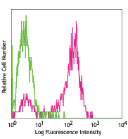
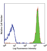
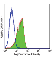
Compare Data Across All Formats
This data display is provided for general comparisons between formats.
Your actual data may vary due to variations in samples, target cells, instruments and their settings, staining conditions, and other factors.
If you need assistance with selecting the best format contact our expert technical support team.


Follow Us