 Login / Register


- Clone
- HCD14 (See other available formats)
- Regulatory Status
- RUO
- Other Names
- LPS receptor
- Isotype
- Mouse IgG1, κ
- Ave. Rating
- Submit a Review
- Product Citations
- publications

-

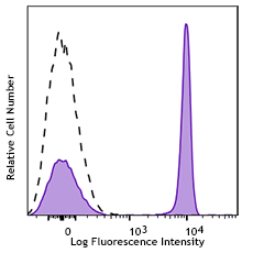
Human peripheral blood monocytes stained with CD14 (clone HCD14) APC/Cyanine7 (filled histogram), or Mouse IgG1, ? APC/Cyanine7 isotype (open histogram).
| Cat # | Size | Price | Quantity Check Availability | Save | ||
|---|---|---|---|---|---|---|
| 325619 | 25 tests | 123€ | ||||
| 325620 | 100 tests | 249€ | ||||
CD14 is a 53-55 kD glycosylphosphatidylinositol (GPI)-linked membrane glycoprotein also known as LPS receptor. CD14 is expressed at high levels on monocytes and macrophages, and at lower levels on granulocytes. Some dendritic cell populations such as interfollicular dendritic cells, reticular dendritic cells, and Langerhans cells have also been reported to express CD14. As a high-affinity receptor for LPS, CD14 is involved in the clearance of gram-negative pathogens and in the upregulation of adhesion molecules and cytokines expression in monocytes and neutrophils.
Product DetailsProduct Details
- Verified Reactivity
- Human
- Antibody Type
- Monoclonal
- Host Species
- Mouse
- Formulation
- Phosphate-buffered solution, pH 7.2, containing 0.09% sodium azide and BSA (origin USA)
- Preparation
- The antibody was purified by affinity chromatography, and conjugated with APC/Cyanine7 under optimal conditions.
- Concentration
- Lot-specific (to obtain lot-specific concentration and expiration, please enter the lot number in our Certificate of Analysis online tool.)
- Storage & Handling
- The antibody solution should be stored undiluted between 2°C and 8°C, and protected from prolonged exposure to light. Do not freeze.
- Application
-
FC - Quality tested
- Recommended Usage
-
Each lot of this antibody is quality control tested by immunofluorescent staining with flow cytometric analysis. For flow cytometric staining, the suggested use of this reagent is 5 µl per million cells in 100 µl staining volume or 5 µl per 100 µl of whole blood.
- Excitation Laser
-
Red Laser (633 nm)
- Application Notes
-
Additional reported applications (for the relevant formats) include: immunofluorescence microscopy. This clone was tested in-house and does not work on formalin fixed paraffin-embedded (FFPE) tissue.
- Additional Product Notes
- BioLegend is in the process of converting the name APC/Cy7 to APC/Cyanine7. The dye molecule remains the same, so you should expect the same quality and performance from our APC/Cyanine7 products. Please contact Technical Service if you have any questions.
- Application References
-
- McMichael A, et al. 1987. Leucocyte Typing III. Oxford University Press. New York.
- Knapp W, et al. Eds. 1989. Leucocyte Typing IV. Oxford University Press. New York.
- Schlossman S, et al. Eds. 1995. Leucocyte Typing V. Oxford University Press. New York.
- Product Citations
-
- RRID
-
AB_830692 (BioLegend Cat. No. 325619)
AB_830693 (BioLegend Cat. No. 325620)
Antigen Details
- Structure
- GPI-linked membrane glycoprotein, 53-55 kD
- Distribution
-
Monocytes, macrophages, granulocytes (low)
- Function
- LPS receptor, clearance of Gram-negative pathogens
- Ligand/Receptor
- LPS
- Cell Type
- Granulocytes, Macrophages, Monocytes, Neutrophils
- Biology Area
- Cell Biology, Immunology, Innate Immunity, Neuroinflammation, Neuroscience
- Molecular Family
- CD Molecules
- Antigen References
-
1. Stocks S, et al. 1990. Biochem. J. 268:275.
2. Wright S, et al. 1990. Science 249:1434. - Gene ID
- 929 View all products for this Gene ID
- Specificity (DOES NOT SHOW ON TDS):
- CD14
- Specificity Alt (DOES NOT SHOW ON TDS):
- CD14
- App Abbreviation (DOES NOT SHOW ON TDS):
- FC
- UniProt
- View information about CD14 on UniProt.org
Related Pages & Pathways
Pathways


Related FAQs
Other Formats
View All CD14 Reagents Request Custom Conjugation| Description | Clone | Applications |
|---|---|---|
| PerCP anti-human CD14 | HCD14 | FC |
| Purified anti-human CD14 | HCD14 | FC,ICC |
| FITC anti-human CD14 | HCD14 | FC |
| PE anti-human CD14 | HCD14 | FC |
| APC anti-human CD14 | HCD14 | FC |
| Alexa Fluor® 488 anti-human CD14 | HCD14 | FC |
| Alexa Fluor® 647 anti-human CD14 | HCD14 | FC,ICC |
| Alexa Fluor® 700 anti-human CD14 | HCD14 | FC |
| Pacific Blue™ anti-human CD14 | HCD14 | FC |
| PE/Cyanine7 anti-human CD14 | HCD14 | FC |
| APC/Cyanine7 anti-human CD14 | HCD14 | FC |
| PerCP/Cyanine5.5 anti-human CD14 | HCD14 | FC |
| Biotin anti-human CD14 | HCD14 | FC,Cell Sep - Pos |
| Brilliant Violet 421™ anti-human CD14 | HCD14 | FC |
| Alexa Fluor® 594 anti-human CD14 | HCD14 | ICC,FC |
| PE/Dazzle™ 594 anti-human CD14 | HCD14 | FC |
| Spark Blue™ 574 anti-human CD14 | HCD14 | FC |
| Brilliant Violet 510™ anti-human CD14 | HCD14 | FC |
| Brilliant Violet 605™ anti-human CD14 | HCD14 | FC |
Customers Also Purchased
Compare Data Across All Formats
This data display is provided for general comparisons between formats.
Your actual data may vary due to variations in samples, target cells, instruments and their settings, staining conditions, and other factors.
If you need assistance with selecting the best format contact our expert technical support team.
-
PerCP anti-human CD14

Human peripheral blood monocytes stained with CD14 (clone HC... -
Purified anti-human CD14

Human peripheral blood monocytes stained with purified HCD14... -
FITC anti-human CD14

Human peripheral blood monocytes stained with HCD14 FITC -
PE anti-human CD14

Human peripheral blood monocytes stained with HCD14 PE 
Pre-lysed human blood leukocytes were stained with PE anti-h... -
APC anti-human CD14

Human peripheral blood monocytes stained with HCD14 APC -
Alexa Fluor® 488 anti-human CD14

Human peripheral blood monocytes stained with HCD14 Alexa Fl... -
Alexa Fluor® 647 anti-human CD14

Human peripheral blood monocytes stained with HCD14 alexa Fl... 
Human peripheral blood mononuclear cells were fixed with 2% ... 
Human peripheral blood mononuclear cells were fixed with 2% ... -
Alexa Fluor® 700 anti-human CD14

Human peripheral blood monocytes stained with HCD14 Alexa Fl... -
Pacific Blue™ anti-human CD14

Human peripheral blood monocytes were stained with CD14 (HCD... -
PE/Cyanine7 anti-human CD14

Human peripheral blood monocytes stained with HCD14 PE/Cyani... -
APC/Cyanine7 anti-human CD14

Human peripheral blood monocytes stained with CD14 (clone HC... -
PerCP/Cyanine5.5 anti-human CD14

Human peripheral blood monocytes were stained with CD14 (Clo... -
Biotin anti-human CD14

Human peripheral blood monocytes stained with biotinylated H... -
Brilliant Violet 421™ anti-human CD14

Human peripheral blood monocytes were stained with CD14 (clo... -
Alexa Fluor® 594 anti-human CD14

Human peripheral blood monocytes were stained with CD14 (clo... 
Human peripheral mononuclear cells were fixed with 2% parafo... -
PE/Dazzle™ 594 anti-human CD14

Human peripheral blood monocytes were stained with CD14 (clo... -
Spark Blue™ 574 anti-human CD14

Human peripheral monocytes were stained with anti-human CD14... -
Brilliant Violet 510™ anti-human CD14

Human peripheral blood monocytes stained with anti-human CD1... 
Human peripheral blood monocytes were stained with anti-huma... -
Brilliant Violet 605™ anti-human CD14

Human peripheral blood monocytes stained with anti-human CD1...


Follow Us