 Login / Register


- Clone
- CD28.2 (See other available formats)
- Regulatory Status
- RUO
- Workshop
- V-CD28.05
- Other Names
- T44, Tp44
- Isotype
- Mouse IgG1, κ
- Ave. Rating
- Submit a Review
- Product Citations
- publications

-

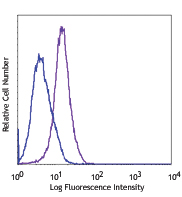
Human peripheral blood lymphocytes stained with APC CD28.2
| Cat # | Size | Price | Quantity Check Availability | Save | ||
|---|---|---|---|---|---|---|
| 302911 | 25 tests | 49€ | ||||
| 302912 | 100 tests | 111€ | ||||
CD28 is a 44 kD disulfide-linked homodimeric type I glycoprotein. It is a member of the immunoglobulin superfamily and is also known as T44 or Tp44. CD28 is expressed on most T lineage cells, NK cell subsets, and plasma cells. CD28 binds both CD80 and CD86 using a highly conserved motif MYPPY in the CDR3-like loop. CD28 is considered a major co-stimulatory molecule, inducing T lymphocyte activation and IL-2 synthesis, and preventing cell death. In vitro studies indicate that ligation of CD28 on T cells by CD80 and CD86 on antigen presenting cells provides a costimulatory signal required for T cell activation and proliferation.
Product DetailsProduct Details
- Reactivity
- Human,Cynomolgus,Rhesus
- Antibody Type
- Monoclonal
- Host Species
- Mouse
- Formulation
- Phosphate-buffered solution, pH 7.2, containing 0.09% sodium azide and BSA (origin USA)
- Preparation
- The antibody was purified by affinity chromatography, and conjugated with APC under optimal conditions.
- Concentration
- Lot-specific (to obtain lot-specific concentration and expiration, please enter the lot number in our Certificate of Analysis online tool.)
- Storage & Handling
- The antibody solution should be stored undiluted between 2°C and 8°C, and protected from prolonged exposure to light. Do not freeze.
- Application
-
FC - Quality tested
- Recommended Usage
-
Each lot of this antibody is quality control tested by immunofluorescent staining with flow cytometric analysis. For flow cytometric staining, the suggested use of this reagent is 5 µl per million cells in 100 µl staining volume or 5 µl per 100 µl of whole blood.
- Excitation Laser
-
Red Laser (633 nm)
- Application Notes
-
The Ultra-LEAF™ Purified antibody (Endotoxin < 0.01 EU/µg, Azide-Free, 0.2 µm filtered) is recommended for highly sensitive assays.
- Application References
-
- Schlossman S, et al. Eds. 1995. Leucocyte Typing V. Oxford University Press. New York.
- Nunes J, et al. 1993. Biochem. J. 293:835.
- Calea-Lauri J, et al. 1999. J. Immunol. 163:62.
- Tazi A, et al. 1999. J. Immunol. 163:3511. (IHC)
- Marti F, et al. 2001. J. Immunol. 166:197. (Costim)
- Jeong SH, et al. 2004. J. Virol. 78:6995. (Costim)
- Rivollier A, et al. 2004. Blood 104:4029. (Costim)
- Scharschmidt E, et al. 2004. Mol. Cell Biol. 24:3860. (Costim)
- Sheng W, et al. 2007.Elsevier 580:6819. PubMed
- Mitsuhashi M. 2007. Clin Chem.53:148. PubMed
- Ye Z, et al. 2008. Infect. Immun. 76:2541. PubMed
- Magatti M, et al. 2008. Stem Cells 26:182. (FA) PubMed
- Yoshino N, et al. 2008. Exp. Anim. (Tokyo) 49:97. (FC)
- Berg M, et al. 2008. J Leukoc Biol. 83:853. (IP) PubMed
- Rout N, et al. 2010. PLoS One 5:e9787. (FC)
- Leonard JA, et al. 2011. J. Virol. 85:6867. PubMed
- Nomura T, et al. 2012. J. Virol. 86:6481. PubMed
- Product Citations
- RRID
-
AB_314313 (BioLegend Cat. No. 302911)
AB_314314 (BioLegend Cat. No. 302912)
Antigen Details
- Structure
- Ig superfamily, type I transmembrane glycoprotein, homodimer, 44 kD
- Distribution
-
Mature T cells, thymocytes, NK cell subsets, plasma cells, EBV-positive B cells
- Function
- T cell costimulation
- Ligand/Receptor
- CD80, CD86
- Cell Type
- B cells, NK cells, Plasma cells, T cells, Thymocytes, Tregs
- Biology Area
- Costimulatory Molecules, Immunology
- Molecular Family
- CD Molecules
- Antigen References
-
1. Schlossman S, et al. Eds. 1995. Leucocyte Typing V. Oxford University Press. New York.
2. June CH, et al. 1994. Immunol. Today 15:321.
3. Linskey PS, et al. 1993. Annu. Rev. Immunol. 11:191. - Gene ID
- 940 View all products for this Gene ID
- UniProt
- View information about CD28 on UniProt.org
Related Pages & Pathways
Pathways


Customers Also Purchased
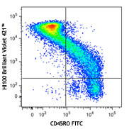
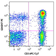
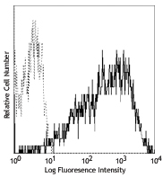
Compare Data Across All Formats
This data display is provided for general comparisons between formats.
Your actual data may vary due to variations in samples, target cells, instruments and their settings, staining conditions, and other factors.
If you need assistance with selecting the best format contact our expert technical support team.


Follow Us