 Login / Register


- Clone
- TER-119 (See other available formats)
- Regulatory Status
- RUO
- Other Names
- Ly-76
- Isotype
- Rat IgG2b, κ
- Ave. Rating
- Submit a Review
- Product Citations
- publications

-

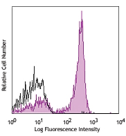
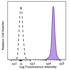
C57BL/6 bone marrow cells were stained with TER-119 APC/Cyanine7 (filled histogram) or Rat IgG2b, κ APC/Cyanine7 isotype control (open histogram). Data shown was gated on small scatter population.
| Cat # | Size | Price | Quantity Check Availability | Save | ||
|---|---|---|---|---|---|---|
| 116223 | 25 µg | 76€ | ||||
The TER-119 antigen is a 52 kD glycophorin A-associated protein, also known as Ly-76. TER-119 is an erythroid-specific antigen expressed on early proerythroblasts to mature erythrocytes, but not on erythroid colony-forming cells (BFU-E, blast-forming unit erythroid, or CFU-E, colony-forming unit erythroid).
Product DetailsProduct Details
- Reactivity
- Mouse
- Antibody Type
- Monoclonal
- Host Species
- Rat
- Immunogen
- Day-14 fetal liver cells from a C57BL/6 mouse
- Formulation
- Phosphate-buffered solution, pH 7.2, containing 0.09% sodium azide.
- Preparation
- The antibody was purified by affinity chromatography, and conjugated with APC/Cyanine7 under optimal conditions.
- Concentration
- 0.2 mg/ml
- Storage & Handling
- The antibody solution should be stored undiluted between 2°C and 8°C, and protected from prolonged exposure to light. Do not freeze.
- Application
-
FC - Quality tested
- Recommended Usage
-
Each lot of this antibody is quality control tested by immunofluorescent staining with flow cytometric analysis. For flow cytometric staining, the suggested use of this reagent is = 0.25 µg per 106 cells in 100 µl volume. It is recommended that the reagent be titrated for optimal performance for each application.
- Excitation Laser
-
Red Laser (633 nm)
- Application Notes
-
The TER-119 antibody is useful for distinguishing erythrocytes and cells in the erythroid lineage. Additional reported applications (for the relevant formats) include: immunoprecipitation1, Western blotting1, complement-mediated cytotoxicity3, and immunohistochemical staining of acetone-fixed frozen sections and formalin-fixed paraffin-embedded sections. Ultra-LEAF™ purified antibody (Endotoxin < 0.01 EU/µg, Azide-Free, 0.2 µm filtered) is recommended for functional assays (Cat. No. 116253-116258).
- Additional Product Notes
- BioLegend is in the process of converting the name APC/Cy7 to APC/Cyanine7. The dye molecule remains the same, so you should expect the same quality and performance from our APC/Cyanine7 products. Please contact Technical Service if you have any questions.
- Application References
-
- Kina T, et al. 2000. Br. J. Haematol. 109:280. (IP, WB)
- Vannucchi AM, et al. 2000. Blood 95:2559.
- Maraskovsky E, et al. 1996. J. Exp. Med. 184:1953. (CMCD)
- Grisendi S, et al. 2005. Nature 437:147. (FC)
- Bourdeau A, et al. 2007. Blood 109:4220.
- Chappaz S, et al. 2007. Blood 110:3862. (FC)
- Heuser M, et al. 2007. Blood 110:1639. (FC)
- Gough SM, et al. 2014. Cancer Discov. 4:564. PubMed
- Product Citations
- RRID
-
AB_2137788 (BioLegend Cat. No. 116223)
Antigen Details
- Structure
- Associated with glycophorin A, 52 kD
- Distribution
-
Early proerythroblast to mature erythrocyte, but not BFU-E and CFU-E
- Cell Type
- Erythrocytes
- Biology Area
- Immunology
- Antigen References
-
1. Kina T, et al. 2000. Br. J. Haematol. 109:280.
2. Ikuta K, et al. 1990. Cell 62:863.
3. Osawa M, et al. 1996. Weir's Handbook of Experimental Immunology. Vol. 2 pp. 66.1-66.5. - Gene ID
- 104231 View all products for this Gene ID
- UniProt
- View information about TER-119 on UniProt.org


Customers Also Purchased
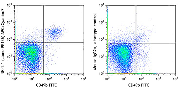
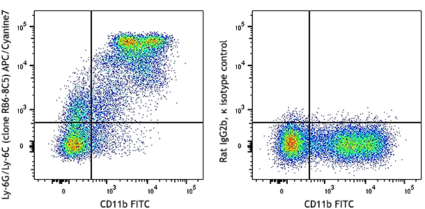
Compare Data Across All Formats
This data display is provided for general comparisons between formats.
Your actual data may vary due to variations in samples, target cells, instruments and their settings, staining conditions, and other factors.
If you need assistance with selecting the best format contact our expert technical support team.


Follow Us