 Login / Register


- Clone
- 63D3 (See other available formats)
- Regulatory Status
- RUO
- Other Names
- Monocyte differentiation antigen CD14, myeloid cell-specific leucine-rich glycoprotein, LPS receptor
- Isotype
- Mouse IgG1, κ
- Ave. Rating
- Submit a Review
- Product Citations
- publications

-

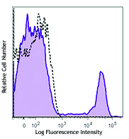
Human peripheral blood monocytes were stained with FITC anti-human CD14 (clone 63D3) (filled histogram) or mouse IgG1, κ FITC isotype control (open histogram).
| Cat # | Size | Price | Quantity Check Availability | Save | ||
|---|---|---|---|---|---|---|
| 367115 | 25 tests | 72€ | ||||
| 367116 | 100 tests | 165€ | ||||
CD14 is a 53-55 kD glycosylphosphatidylinositol (GPI)-linked membrane glycoprotein that is also known as the LPS receptor. CD14 is expressed at high levels on monocytes and macrophages, and at lower levels on granulocytes. Some dendritic cell populations such as interfollicular dendritic cells, reticular dendritic cells, and Langerhans cells have also been reported to express CD14. As a high-affinity receptor for LPS, CD14 is involved in the clearance of gram-negative pathogens and in the upregulation of adhesion molecules and cytokine expression in monocytes and neutrophils.
Product DetailsProduct Details
- Reactivity
- Human
- Antibody Type
- Monoclonal
- Host Species
- Mouse
- Immunogen
- Purified human peripheral blood monocytes.
- Formulation
- Phosphate-buffered solution, pH 7.2, containing 0.09% sodium azide and BSA (origin USA)
- Preparation
- The antibody was purified by affinity chromatography and conjugated with FITC under optimal conditions.
- Concentration
- Lot-specific (to obtain lot-specific concentration and expiration, please enter the lot number in our Certificate of Analysis online tool.)
- Storage & Handling
- The antibody solution should be stored undiluted between 2°C and 8°C, and protected from prolonged exposure to light. Do not freeze.
- Application
-
FC - Quality tested
- Recommended Usage
-
Each lot of this antibody is quality control tested by immunofluorescent staining with flow cytometric analysis. For flow cytometric staining, the suggested use of this reagent is 5 µl per million cells in 100 µl staining volume or 5 µl per 100 µl of whole blood.
- Excitation Laser
-
Blue Laser (488 nm)
- Application References
-
- Fridlender ZG, et al. 1999. Hum. Immunol. 11:1028.
- Devitt A, et al. 1998. Nature 6675:505.
- Product Citations
- RRID
-
AB_2571928 (BioLegend Cat. No. 367115)
AB_2571929 (BioLegend Cat. No. 367116)
Antigen Details
- Structure
- GPI-linked membrane glycoprotein.
- Distribution
-
Monocytes, macrophages, dendritic cells, and granulocytes.
- Function
- LPS receptor, clearance of Gram-negative pathogens.
- Interaction
- LPS.
- Ligand/Receptor
- LPS.
- Cell Type
- Dendritic cells, Granulocytes, Macrophages, Monocytes, Neutrophils
- Biology Area
- Cell Biology, Immunology, Neuroinflammation, Neuroscience
- Molecular Family
- Adhesion Molecules, CD Molecules
- Antigen References
-
- Stocks SC, et al. 1990. Biochem. J. 268:275.
- Wright SD, et al. 1990. Science 4975:1431.
- Gene ID
- 929 View all products for this Gene ID
- UniProt
- View information about CD14 on UniProt.org
Related Pages & Pathways
Pathways


Customers Also Purchased
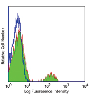
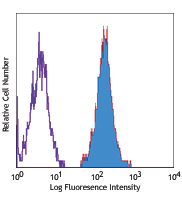
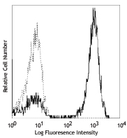
Compare Data Across All Formats
This data display is provided for general comparisons between formats.
Your actual data may vary due to variations in samples, target cells, instruments and their settings, staining conditions, and other factors.
If you need assistance with selecting the best format contact our expert technical support team.


Follow Us