 Login / Register


- Clone
- 2H7 (See other available formats)
- Regulatory Status
- RUO
- Workshop
- IV B201
- Other Names
- B1, Bp35
- Isotype
- Mouse IgG2b, κ
- Ave. Rating
- Submit a Review
- Product Citations
- publications

-

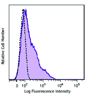
Human peripheral blood lymphocytes were stained with anti-CD20 (clone 2H7) Pacific Blue™ (filled histogram), or mouse IgG2b, κ Pacific Blue™ (open histogram).
CD20 is a 33-37 kD, four transmembrane spanning protein, also known as B1 and Bp35. CD20 is expressed on pre-B-cells, resting and activated B cells (not plasma cells), some follicular dendritic cells, and at low levels on a T cell subset. CD20 is heavily phosphorylated on activated B cells and malignant B cells. Homo-oligomeric complexes of CD20 are thought to form Ca2+ conductive ion channels in the plasma membrane of B cells. The CD20 molecule is involved in B-cell activation and is associated with various Src family kinases (Lyn, Lck, Fyn). It exists in a complex with MHC class I and II, CD53, CD81, and CD82.
Product DetailsProduct Details
- Reactivity
- Human,Cynomolgus,Rhesus
- Antibody Type
- Monoclonal
- Host Species
- Mouse
- Immunogen
- Human tonsillar B cells
- Formulation
-
test size: Phosphate-buffered solution, pH 7.2, containing 0.09% sodium azide and BSA (origin USA).
µg sizes: Phosphate-buffered solution, pH 7.2, containing 0.09% sodium azide. - Preparation
- The antibody was purified by affinity chromatography, and conjugated with Pacific Blue™ under optimal conditions.
- Concentration
- µg sizes: 0.5 mg/mLtest sizes: lot-specific (to obtain lot-specific concentration and expiration, please enter the lot number in our Certificate of Analysis online tool.)
- Storage & Handling
- The antibody solution should be stored undiluted between 2°C and 8°C, and protected from prolonged exposure to light. Do not freeze.
- Application
-
FC - Quality tested
- Recommended Usage
-
Each lot of this antibody is quality control tested by immunofluorescent staining with flow cytometric analysis.
For test size, the suggested use of this reagent for immunofluorescent staining is 5 µl per 106 cells in 100 µl volume.
For µg sizes, the suggested use of this reagent for immunofluorescent staining is ≤0.5 µg per 106 cells in 100 µl volume.
It is recommended that the reagent be titrated for optimal performance for each application.
* Pacific Blue™ has a maximum emission of 455 nm when it is excited at 405 nm. Prior to using Pacific Blue™ conjugate for flow cytometric analysis, please verify your flow cytometer's capability of exciting and detecting the fluorochrome.
Alexa Fluor® and Pacific Blue™ are trademarks of Life Technologies Corporation.
View full statement regarding label licenses - Excitation Laser
-
Violet Laser (405 nm)
- Application Notes
-
The epitope recognized by clone 2H7 has been mapped to the sequence YNCEPANPSEKNSPST which lies in the large extracellular loop of human CD20. Additional reported applications (for the relevant formats) include: immunoprecipitation4 and immunohistochemical staining of acetone-fixed frozen sections5.
- Application References
-
- Schlossman S, et al. 1995. Leucocyte Typing V. Oxford University Press. New York.
- Knapp W, et al. 1989. Leucocyte Typing IV. Oxford University Press. New York.
- McMichael A, et al. Eds. 1987. Leucocyte Typing III Oxford University Press. New York.
- Polyak MJ, et al. 2002. Blood 99:3256. (IP)
- Mack CL, et al. 2004. Pediatr. Res. 56:79. (IHC)
- Product Citations
- RRID
-
AB_493650 (BioLegend Cat. No. 302319)
AB_493651 (BioLegend Cat. No. 302320)
AB_1595435 (BioLegend Cat. No. 302328)
Antigen Details
- Structure
- Four transmembrane protein (TM4SF), heavily phosphorylated after activation, 33-37 kD
- Distribution
-
B cell, T cell subsets
- Function
- B cell activation
- Ligand/Receptor
- Src family tyrosine kinases, MHC class I, II, CD53, CD81, CD82
- Cell Type
- B cells, T cells
- Biology Area
- Costimulatory Molecules, Immunology
- Molecular Family
- CD Molecules
- Antigen References
-
1. Hultin L, et al. 1993. Cytometry 14:196.
2. Tedder T, et al. 1994. Immunol. Today 15:450. - Gene ID
- 931 View all products for this Gene ID
- UniProt
- View information about CD20 on UniProt.org
Related Pages & Pathways
Pathways


Related FAQs
- What is the difference between two anti human CD20 clones 2H7 and 1412?
-
For clone 1412: This clone specifically recognizes cytoplasmic domain of CD20 and thus can only be used for intracellular flow cytometry. In this instance you will need to include the fixation and permeabilization steps. Please follow the intracellular flow cytometry staining protocol.
For clone 2H7: This clone is ok for regular surface staining for CD20 and there is no need for any fixation and permeabilization steps.
Our technical protocols can be found here.
Customers Also Purchased
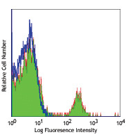
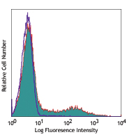
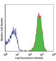
Compare Data Across All Formats
This data display is provided for general comparisons between formats.
Your actual data may vary due to variations in samples, target cells, instruments and their settings, staining conditions, and other factors.
If you need assistance with selecting the best format contact our expert technical support team.


Follow Us