 Login / Register


- Clone
- SK7 (See other available formats)
- Regulatory Status
- RUO
- Workshop
- HCDM listed
- Other Names
- T3, CD3ε
- Isotype
- Mouse IgG1, κ
- Ave. Rating
- Submit a Review
- Product Citations
- publications

-

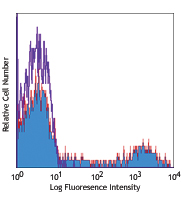
Human peripheral blood lymphocytes stained with SK7 PerCP
| Cat # | Size | Price | Quantity Check Availability | Save | ||
|---|---|---|---|---|---|---|
| 344813 | 25 tests | 115€ | ||||
| 344814 | 100 tests | 252€ | ||||
CD3ε is a 20 kD chain of the CD3/T-cell receptor (TCR) complex, which is composed of two CD3ε, one CD3γ, one CD3δ, one CD3ζ (CD247), and a T-cell receptor (α/β or γ/δ) heterodimer. It is found on all mature T cells, NK T cells, and some thymocytes. CD3, also known as T3, is a member of the immunoglobulin superfamily that plays a role in antigen recognition, signal transduction, and T cell activation.
Product DetailsProduct Details
- Reactivity
- Human
- Antibody Type
- Monoclonal
- Host Species
- Mouse
- Formulation
- Phosphate-buffered solution, pH 7.2, containing 0.09% sodium azide and BSA (origin USA)
- Preparation
- The antibody was purified by affinity chromatography, and conjugated with PerCP under optimal conditions.
- Concentration
- Lot-specific (to obtain lot-specific concentration and expiration, please enter the lot number in our Certificate of Analysis online tool.)
- Storage & Handling
- The antibody solution should be stored undiluted between 2°C and 8°C, and protected from prolonged exposure to light. Do not freeze.
- Application
-
FC - Quality tested
- Recommended Usage
-
Each lot of this antibody is quality control tested by immunofluorescent staining with flow cytometric analysis. For flow cytometric staining, the suggested use of this reagent is 5 µl per million cells in 100 µl staining volume or 5 µl per 100 µl of whole blood.
- Excitation Laser
-
Blue Laser (488 nm)
- Application Notes
-
Additional reported application (for the relevant formats) include: immunohistochemical staining of frozen tissue sections4,5,8, immunofluorescent staining6, and Western blotting3.
- Application References
-
- Kan EA, et al. 1983. J. Immunol. 131:536.
- Wood GS, et al. 1985. Am. J. Pathol. 120:371.
- Van Dongen JJM, et al. 1988. Blood 71:603. (WB)
- Haringman JJ, et al. 2005. Arthritis Res. Ther. 7:R862. (IHC)
- Carbone A, et al. 1999. Blood 93:2319. (IHC)
- Goval JJ, et al. 2006. J. Histochem. Cytochem. 54:75. (IF)
- Rutjens E, et al. 2007. J. Immunol. 178:1702.
- Kap Y, et al. 2009. J. Histochem. Cytochem. 57:1159. (IHC)
- Yoshino N, et al. 2000. Exp. Anim. (Tokyo) 49:97. (FC)
- Product Citations
- RRID
-
AB_10641841 (BioLegend Cat. No. 344813)
AB_10639948 (BioLegend Cat. No. 344814)
Antigen Details
- Structure
- Ig superfamily, with the subunits of CD3γ, CD3δ, CD3ζ, (CD247) and TCR (α/β or γ/δ) forms CD3/TCR complex, 20 kD
- Distribution
-
Mature T and NK T cells, during thymocyte differentiation
- Function
- Antigen recognition, signal transduction, T cell activation
- Ligand/Receptor
- Peptide antigen bound to MHC
- Cell Type
- NKT cells, T cells, Tregs
- Biology Area
- Immunology, Innate Immunity
- Molecular Family
- CD Molecules, TCRs
- Antigen References
-
1. Barclay N, et al. 1993. The Leucocyte FactsBook. Academic Press. San Diego.
2. Beverly P, et al. 1981. Eur. J. Immunol. 11:329.
3. Lanier L, et al. 1986. J. Immunol. 137:2501. - Gene ID
- 916 View all products for this Gene ID
- UniProt
- View information about CD3 on UniProt.org
Related Pages & Pathways
Pathways


Related FAQs
- How stable is PerCP/Cy5.5 tandem as compared to PerCP alone?
-
PerCP/Cy5.5 is quite photostable and also better than PerCP alone in withstanding fixation.
Customers Also Purchased
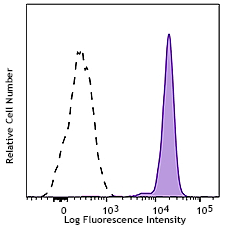
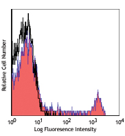
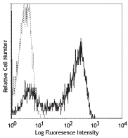
Compare Data Across All Formats
This data display is provided for general comparisons between formats.
Your actual data may vary due to variations in samples, target cells, instruments and their settings, staining conditions, and other factors.
If you need assistance with selecting the best format contact our expert technical support team.


Follow Us