 Login / Register


- Clone
- 2E1.E9 (See other available formats)
- Regulatory Status
- RUO
- Other Names
- Glial fibrillary acid protein
- Isotype
- Mouse IgG2b
- Ave. Rating
- Submit a Review
- Product Citations
- publications

-

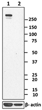
Total cell lysate from HeLa (negative control), U87-MG and tissue lysate from mouse brain (15 µg/lane) were resolved by electrophoresis (4-12% Bis-Tris gel), transferred to nitrocellulose membrane, and probed with 0.2 µg/mL of purified anti-GFAP. Proteins were visualized using a goat anti-mouse-IgG secondary antibody conjugated to HRP and chemiluminescence detection system. Direct-Blot™ HRP anti-β-actin antibody was used as a loading control (lower). Lane M is the molecular weight ladder. -

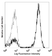
U251 cells were fixed with 4% paraformaldehyde (PFA) for ten minutes, permeabilized with 0.5% Triton X-100 for five minutes and blocked with 5% FBS for 30 minutes. Then, the cells were intracellularly stained with 5 µg/mL anti-GFAP (clone 2E1.E9) antibody followed by Alexa Fluor® 594 Goat anti-mouse IgG (minimal x-reactivity) Antibody (red) for one hour at room temperature. Nuclei were counterstained with DAPI (blue). The image was captured with a 60X objective. -

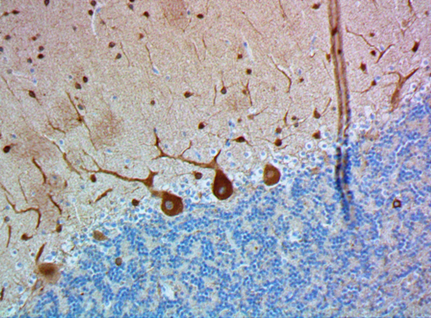
C57BL/6 mouse frozen brain section was fixed with 4% paraformaldehyde (PFA) for 10 minutes at room temperature and blocked with 5% FBS for 30 minutes at room temperature. Then the section was stained with 10 µg/ml of anti-GFAP (clone 2E1.E9) purified overnight at 4°C, followed by 2.5 µg/ml of anti-mouse IgG2b (clone RMG2b-1) Alexa Fluor® 594 (red) for 2 hours at room temperature. Nuclei were counterstained with DAPI (blue). The image was captured by 10X objective. -

IHC staining of Purified anti-GFAP (clone 2E1.E9) on formalin-fixed paraffin-embedded human brain tissue. Following antigen retrieval using Citrate Buffer, 10X (Cat. No. 420902), the tissue was incubated with 5 µg/mL of purified anti-GFAP (clone 2E1.E9) followed by incubation with Alexa Fluor® 647 goat anti-mouse IgG (Cat. No. 405322) for 1 hour at room temperature. Nuclei were counterstained with DAPI (Cat. No. 422801). Images were captured with a 20X (panel A) and 40X (panel B) objective. Scale bar: 25 µm -

IHC staining of purified anti-GFAP (clone 2E1.E9) on formalin-fixed paraffin-embedded human brain tissue. Following antigen retrieval using Tris-EDTA pH 9.0 buffer (10 mM Tris, 1 mM EDTA), the tissue was incubated with 2 µg/mL of purified anti-GFAP (clone 2E1.E9) followed by incubation with Alexa Fluor® 647 goat anti-mouse IgG (Cat. No. 405322) (panel A) for 1 hour at room temperature. Tissue was also stained with 2 µg/mL of Alexa Fluor® 594 anti-Tubulin β 3 (Cat. No. 657408) (green), and nuclei were counterstained with DAPI (Cat. No. 422801) (blue). Merged images (panel B). Images were captured with a 20X objective. Scale bar: 50 µm
| Cat # | Size | Price | Quantity Check Availability | Save | ||
|---|---|---|---|---|---|---|
| 644701 | 25 µg | 100€ | ||||
| 644702 | 100 µg | 235€ | ||||
GFAP is expressed exclusively in astrocytes in the central nervous system. The protein is a member of the intermediate filament family of proteins which form networks providing support and strength to cells. This antibody does not cross-react with other intermediate filaments such as vimentin, neurofilament proteins, desmin and others. More than 50 GFAP mutations have been identified to be associated with the Alexander disease.
Product DetailsProduct Details
- Reactivity
- Human,Mouse,Rat
- Antibody Type
- Monoclonal
- Host Species
- Mouse
- Immunogen
- Bovine spinal cord homogenate
- Formulation
- Phosphate-buffered solution, pH 7.2, containing 0.09% sodium azide.
- Preparation
- The antibody was purified by affinity chromatography.
- Concentration
- 0.5 mg/mL
- Storage & Handling
- The antibody solution should be stored undiluted between 2°C and 8°C.
- Application
-
WB - Quality tested
ICC, IHC-F, IHC-P - Verified
FC - Reported in the literature, not verified in house - Recommended Usage
-
Each lot of this antibody is quality control tested by Western blotting. Western blotting, suggested working dilution(s): Use 0.25 - 1.0 µg antibody per mL antibody dilution buffer for each mini-gel. For immunocytochemistry, a concentration range of 1.0 - 5.0 µg/mL is recommended. For immunohistochemical staining of frozen tissue sections, a concentration range of 5.0 - 10 µg/mL is suggested. For immunohistochemistry on formalin-fixed paraffin-embedded tissue sections, a concentration range of 1.0 - 10 µg/mL is suggested. It is recommended that the reagent be titrated for optimal performance for each application.
- Application Notes
-
For Immunohistochemistry, Citrate buffer, 10X (Cat. No. 420902) or Tris-EDTA pH 9.0 is recommended.
- Application References
-
- McLendon RE and Bigner DD. 1994. Brain Pathol. 4:221.
- Liu W, et al. 2011. Proteomics 11:3556. (FC) PubMed
- Product Citations
- RRID
-
AB_2109791 (BioLegend Cat. No. 644701)
AB_2294566 (BioLegend Cat. No. 644702)
Antigen Details
- Structure
- Around 50 kD
- Distribution
-
Only expressed in astrocytes in the central nervous system.
- Function
- Together with other proteins to form intermediate filaments which supports astroglial cells. Astroglial cells support and nourish cells in the brain and spinal cord. If brain cells are injured through trauma or disease, astroglial cells react by rapidly prodcing more GFAP protein.
- Cell Type
- Astrocytes
- Biology Area
- Cell Biology, Neuroscience, Neuroscience Cell Markers
- Molecular Family
- Intermediate Filaments
- Gene ID
- 2670 View all products for this Gene ID
- UniProt
- View information about GFAP on UniProt.org

Customers Also Purchased
Compare Data Across All Formats
This data display is provided for general comparisons between formats.
Your actual data may vary due to variations in samples, target cells, instruments and their settings, staining conditions, and other factors.
If you need assistance with selecting the best format contact our expert technical support team.


Follow Us