 Login / Register


- Clone
- PK136 (See other available formats)
- Regulatory Status
- RUO
- Other Names
- NKR-P1C, NKR-P1B, Ly-55, CD161, CD161b, CD161c
- Isotype
- Mouse IgG2a, κ
- Ave. Rating
- Submit a Review
- Product Citations
- publications

-

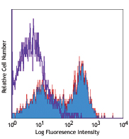
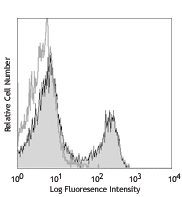
C57BL/6 mouse splenocytes were stained with CD49b (DX5) APC and purified NK1.1 (clone PK136) (left) or purified mouse IgG2a, κ isotype control (right), followed by anti-mouse IgG2a PE.
Select size of product is eligible for a 40% discount! Promotion valid until September 30, 2024. Exclusions apply. To view full promotion terms and conditions or to contact your local BioLegend representative to receive a quote, visit our webpage.
NK-1.1 surface antigen, also known as CD161b/CD161c and Ly-55, is encoded by the NKR-P1B/NKR-P1C gene. It is expressed on NK cells and NK-T cells in some mouse strains, including C57BL/6, FVB/N, and NZB, but not AKR, BALB/c, CBA/J, C3H, DBA/1, DBA/2, NOD, SJL, and 129. Expression of NKR-P1C antigen has been correlated with lysis of tumor cells in vitro and rejection of bone marrow allografts in vivo. NK-1.1 has also been shown to play a role in NK cell activation, IFN-γ production, and cytotoxic granule release. NK-1.1 and DX5 are commonly used as mouse NK cell markers.
Product DetailsProduct Details
- Reactivity
- Mouse
- Antibody Type
- Monoclonal
- Host Species
- Mouse
- Immunogen
- NK-1+ cells from mouse spleen and bone marrow
- Formulation
- 0.2 µm filtered in phosphate-buffered solution, pH 7.2, containing no preservative.
- Preparation
- The Ultra-LEAF™ (Low Endotoxin, Azide-Free) antibody was purified by affinity chromatography.
- Concentration
- The antibody is bottled at the concentration indicated on the vial, typically between 2 mg/mL and 3 mg/mL. Older lots may have also been bottled at 1 mg/mL. To obtain lot-specific concentration and expiration, please enter the lot number in our Certificate of Analysis online tool.
- Storage & Handling
- The antibody solution should be stored undiluted between 2°C and 8°C. This Ultra-LEAF™ solution contains no preservative; handle under aseptic conditions.
- Application
-
FC - Quality tested
CyTOF® - Verified
IP, Deplete, Block, IHC-F - Reported in the literature, not verified in house - Recommended Usage
-
Each lot of this antibody is quality control tested by immunofluorescent staining with flow cytometric analysis. For flow cytometric staining, the suggested use of this reagent is ≤ 0.25 µg per million cells in 100 µl volume. It is recommended that the reagent be titrated for optimal performance for each application.
- Application Notes
-
Additional reported applications (for the relevant formats) include: immunoprecipitation1,2, complement-dependent cytotoxicity3, in vivo depletion4,5,9,10, mediation of in vitro redirected lysis6, blocking of NK cell function7, induction of proliferation8, immunohistochemical staining of frozen sections11, immunofluorescence microscopy11, and spatial biology (IBEX)16,17. The LEAF™ purified antibody (Endotoxin <0.1 EU/µg, Azide-Free, 0.2 µm filtered) is recommended for functional assays (Cat. No. 108712).
- Application References
-
- Carlyle JR, et al. 1999. J. Immunol. 162:5917. (IP)
- Sentman CL, et al. 1989. Hybridoma 8:605. (IP)
- Koo GC, et al. 1984. Hybridoma 3:301. (Cyt)
- Sentman CL, et al. 1989. J. Immunol. 142:1847. (Deplete)
- Koo GC, et al. 1986. J. Immunol. 137:3742. (Deplete)
- Karlhofer FM, et al. 1991. J. Immunol. 146:3662.
- Kung SK, et al. 1999. J. Immunol. 162:5876. (Block)
- Reichlin A, et al. 1998. Immunol. Cell Biol. 76:143.
- Drobyski W, et al. 1996. Blood 87:5355. (Deplete)
- Andoniou CE, et al. 2005. Nat. Immunol. 6:1011. (Deplete)
- Kanwar JR, et al. 2001. J. Natl. Cancer Inst. 93:1541. (IHC, IF)
- Kroemer A, et al. 2008. J. Immunol. 180:7818. PubMed
- Kim JY, et al. 2009. Exp Mol Med. 30:288. PubMed
- Bankoti J, et al. 2010. Toxicol. Sci. 115:422. (FC) PubMed
- Lee H, et al. 2014. Invest Ophthalmol Vis Sci. 55:2885. PubMed
- Radtke AJ, et al. 2020. Proc Natl Acad Sci U S A. 117:33455-65. (SB) PubMed
- Radtke AJ, et al. 2022. Nat Protoc. 17:378-401. (SB) PubMed
- Product Citations
- RRID
-
AB_2800571 (BioLegend Cat. No. 108761)
AB_2800572 (BioLegend Cat. No. 108762)
AB_2800570 (BioLegend Cat. No. 108760)
AB_2800567 (BioLegend Cat. No. 108757)
AB_2800568 (BioLegend Cat. No. 108758)
AB_2800569 (BioLegend Cat. No. 108759)
Antigen Details
- Structure
- NKR-P1 gene family
- Distribution
-
NK and NK-T cells in the NK1.1 mouse strains (C57BL, FVB/N, NZB)
- Function
- NK cell activation, IFN-γ production, cytotoxic granule release
- Cell Type
- NK cells, NKT cells
- Biology Area
- Immunology, Innate Immunity
- Antigen References
-
1. Lanier LL. 1997. Immunity 6:371.
2. Yokoyama WM, et al. 1993. Ann. Rev. Immunol. 11:613.
3. Koo GC, et al. 1986. J. Immunol. 137:3742.
4. Giorda R, et al. 1991. J. Immunol. 147:1701. - Gene ID
- 17059 View all products for this Gene ID
- UniProt
- View information about NK-1.1 on UniProt.org


Related FAQs
- Do you guarantee that your antibodies are totally pathogen free?
-
BioLegend does not test for pathogens in-house aside from the GoInVivo™ product line. However, upon request, this can be tested on a custom basis with an outside, independent laboratory.
- Does BioLegend test each Ultra-LEAF™ antibody by functional assay?
-
No, BioLegend does not test Ultra-LEAF™ antibodies by functional assays unless otherwise indicated. Due to the possible complexities and variations of uses of biofunctional antibodies in different assays and because of the large product portfolio, BioLegend does not currently perform functional assays as a routine QC for the antibodies. However, we do provide references in which the antibodies were used for functional assays and we do perform QC to verify the specificity and quality of the antibody based on our strict specification criteria.
- Does BioLegend test each Ultra-LEAF™ antibody for potential pathogens?
-
No, BioLegend does not test for pathogens in-house unless otherwise indicated. However, we can recommend an outside vendor to perform this testing as needed.
- Have you tested this Ultra-LEAF™ antibody for in vivo or in vitro applications?
-
We don't test our antibodies for in vivo or in vitro applications unless otherwise indicated. Depending on the product, the TDS may describe literature supporting usage of a particular product for bioassay. It may be best to further consult the literature to find clone specific information.
Customers Also Purchased
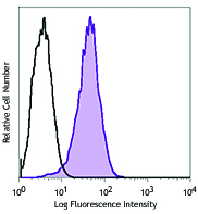
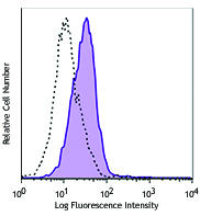
Compare Data Across All Formats
This data display is provided for general comparisons between formats.
Your actual data may vary due to variations in samples, target cells, instruments and their settings, staining conditions, and other factors.
If you need assistance with selecting the best format contact our expert technical support team.


Follow Us