 Login / Register


- Regulatory Status
- RUO
- Other Names
- Please refer to individual product datasheets.
- Ave. Rating
- Submit a Review
- Product Citations
- publications

-

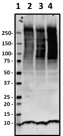
Western blot of anti-Ubiquitin antibody (clone P4G7). Lane 1: Molecular weight marker; Lane 2: Poly-His tagged human ubiquitin protein. The blot was incubated with the primary antibody at 10 µg/ml for 24 hours at 4°C, followed by incubation with HRP labeled goat anti-mouse secondary antibody and visualization with a chemiluminescence detection system. -

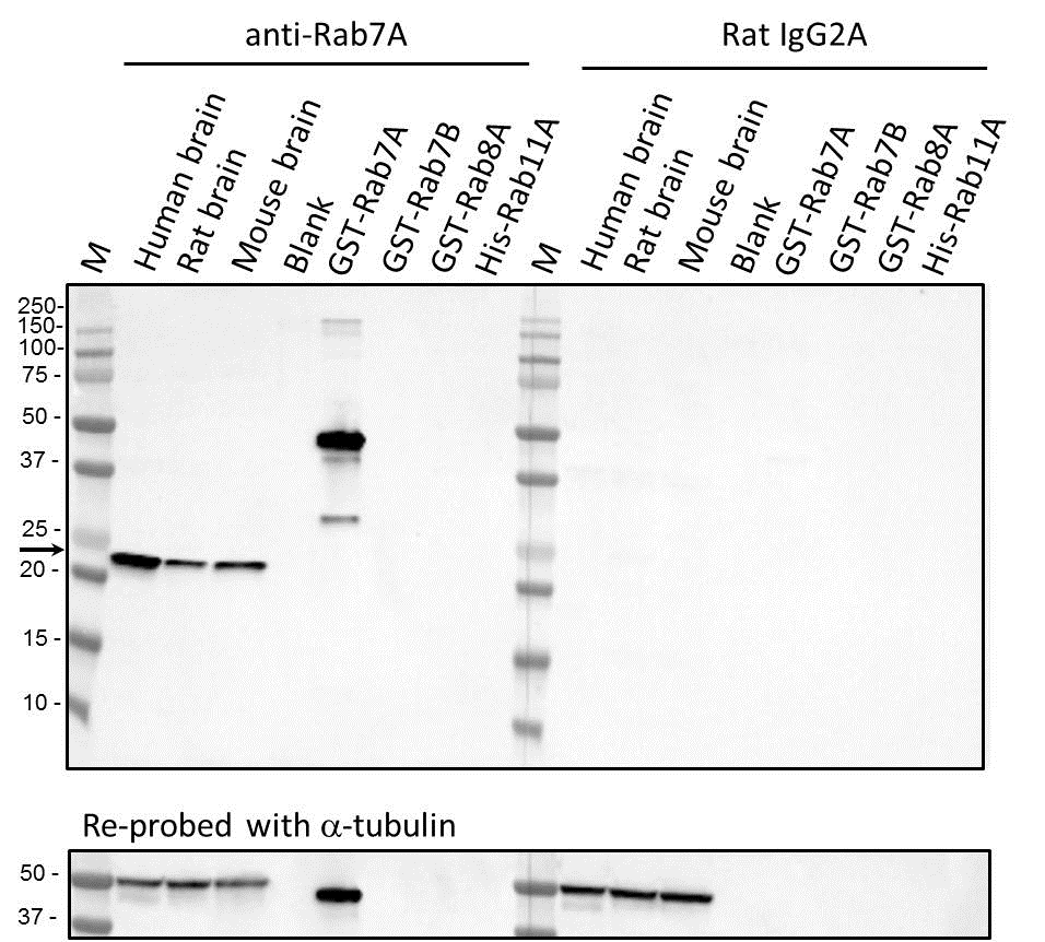
Western blot of anti-Rab7A antibody (clone W16034A). The blot was incubated with 2µg/ml of the primary antibody overnight at 4°C, followed by the incubation with horseradish peroxidase labeled goat anti-mouse secondary antibody. Enhanced chemiluminescence was used as the detection system. M: Molecular weight marker; brain lysates: 20 µg; recombinant proteins: 10 ng. -

Western blot of anti-Beclin-1 antibody (clone O93F3) and isotype-matched IgG1 control. Blots were incubated with 5µg/ml (left) or 0.5µg/ml (middle) anti-Beclin-1 antibody or IgG1 isotype control antibody (right) overnight at 4°C, followed by the incubation with horseradish peroxidase labeled goat anti-mouse secondary antibody. Enhanced chemiluminescence was used as the detection system. M: Molecular weight marker; brain lysates: 20 µg. The anti-Histone-H3 antibody was used as a loading control. -

Western blot of anti-ATG5 antibody (clone 177.19). The blot was incubated with the primary antibody at 0.2 µg/ml overnight at 4°C, followed by the incubation with horseradish peroxidase labeled goat anti-mouse secondary antibody (Cat. No. 405306). Enhanced chemiluminescence was used as the detection system. Lysates: 10 µg. Recombinant human ATG5: 5 ng. -

ICC staining of anti-ATG5 antibody (clone 177.19) on Hela cells. The cells were fixed with 4% PFA, permeabilized with 0.1% Triton X-100, and blocked with 2% normal goat serum and 0.02%BSA. The cells were then stained with 0.5 µg/ml of the primary antibody overnight at 4°C, followed by incubation with Alexa Fluor® 594 goat anti-Mouse IgG (Cat. No. 405326) for one hour at room temperature. Cells were counterstained with Phalloidin™ Green 488 (Cat. No. 424201) and DAPI to visualize actin filaments and nuclei, respectively. The images were captured with a 60X objective. Scale bar: 50 µm. -

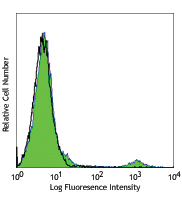
SH-SY5Y neuroblastoma cells were fixed with 4% paraformaldehyde, and then permeabilized with 0.1% Triton x-100 for 20 minutes. Cells were blocked with 2% normal goat serum for 30 minutes at room temperature, and then incubated with anti-LC3 antibody (clone A15143K) at 1 µg/mL for three hours at room temperature. Alexa Fluor® 488 conjugated anti-mouse IgG (green) was used as secondary antibody at 1 µg/mL. Cells were then incubated with anti-Tubulin β3 (TUBB3) antibody (clone TUJ1) Alexa Fluor® 647 (pink) at 1 µg/mL overnight at 4°C. Nuclei were stained with Hoechst 33342 (blue) at 5 µg/mL for 10 minutes at room temperature. Images were captured with a 60X objective.
| Cat # | Size | Price | Quantity Check Availability | Save | ||
|---|---|---|---|---|---|---|
| 899901 | 1 kit | 457 CHF | ||||
Autophagy is a regulated process for protein degradation and turnover of damaged organelles to maintain cellular homeostasis. Under conditions such as stress or starvation, autophagy contributes to the breakdown of proteins and cellular compartments to generate amino acids required for the synthesis of proteins essential for cell survival. Dysregulation of autophagy or mutations in proteins involved in this pathway have been shown to contribute to neurodegenerative disorders such as Parkinson’s disease. The Autophagy Antibody Sampler Kit provides flexibility for sampling and detection of key targets in the autophagy pathway.
Product DetailsKit Contents
- Kit Contents
-
Specificity Format Clone Size Reactivity Isotype Anti-Ubiquitin Purified P4G7 25 μg Human, Extensive (Yeast to Human) Mouse IgG1, κ Anti-Rab7A Purified W16034A 25 μg Human, Rat, Mouse Rat IgG2a, κ Anti-Beclin-1 Purified O93F3 25 μg Human, Rat, Mouse Mouse IgG1 Anti-ATG5 Purified 177.19 25 μg Human, Mouse, Rat Mouse IgG1, κ Anti-LC3 Purified A15143K 25 μg Human, Mouse Mouse IgG2b, κ * For detailed information about each specificity, please refer to the datasheets of the individual products.
Product Details
- Formulation
- Please refer to individual product datasheets of the purified formats for details.
- Preparation
- All antibodies in this kit were purified by affinity chromatography.
- Storage & Handling
- Upon receipt, store undiluted at 2-8°C.
- Application
-
WB, IHC-P - Quality tested
ICC - Verified - Recommended Usage
-
Each lot of antibodies in this kit is quality control tested by Western blotting. For Western blotting, the suggested uses of these reagents are as follows:
Anti-Ubiquitin: 1.0 - 10 µg/ml
Anti-Rab7A: 0.001 - 0.005 µg/ml
Anti-Beclin-1: 0.5 - 5.0 µg/ml
Anti-ATG5: 0.1 - 1.0 µg/ml
For immunohistochemistry, the suggested uses of these reagents are as follows:
Anti-ATG5: 0.2 - 1.0 µg/ml
Anti-LC3: 0.5 - 1.0 µg/ml
It is recommended that the reagent be titrated for optimal performance for each application. - Application Notes
-
For verified or reported applications for these antibodies, please see individual product datasheets.
Antigen Details
- Biology Area
- Cell Biology, Neurodegeneration, Neuroscience, Protein Trafficking and Clearance
- Antigen References
-
- Uchiyama Y, et al. 2008. Histochem. Cell Biol. 129:407.
- Wu H, et al. 2014. Int. J. Biol. Sci. 10:1072.
- Subramani S, Malhotra V. 2013. EMBO Rep. 14(2):143.
- Cao Y, et al. 2007. Cell. Research 17:839.
- Lee J, et al. 2012. Biochem. J. 441(2):523.
- Martini-Stoica H, et al. 2016. Trends Neurosci. 39(4)221.
- Guerra F, Bucci C. 2016. Cells. 5(3).
- Hatanaka H, et al. 2009. J. Biol. Chem. 23:15448.
- Gene ID
- 9474 View all products for this Gene ID 8678 View all products for this Gene ID 81631 View all products for this Gene ID 7879 View all products for this Gene ID 7316 View all products for this Gene ID


Related FAQs
Customers Also Purchased


Follow Us