 Login / Register


- Clone
- UCHT1 (See other available formats)
- Regulatory Status
- RUO
- Workshop
- III 471
- Other Names
- T3, CD3ε
- Isotype
- Mouse IgG1, κ
- Ave. Rating
- Submit a Review
- Product Citations
- publications

-

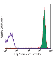
Human peripheral blood lymphocytes stained with UCHT1 Alexa Fluor 488® -

Human peripheral blood mononuclear cells and neutrophil mixed cells were fixed with 2% paraformaldehyde (PFA), and then stained with 5 µg/ml CD11b (clone ICRF44) Alexa Fluor® 594 (red) and 5 µg/ml CD3 (clone UCHT1) Alexa Fluor® 488 (green) for 30 minutes at room temperature. Nuclei were counterstained with DAPI (blue). The image was captured by 40X objective. -

Human frozen tonsil tissue slices were fixed with 4% PFA for ten minutes and blocked with 5% FBS for 30 minutes. Then, the tissue was stained with 5 µg/mL of Alexa Fluor® 488 anti-human CD3 antibody (green) and Brilliant Violet 421™ anti-human CD19 antibody (blue) overnight at 4°C. The image was scanned with a 10X objective and stitched with MetaMorph® software.
| Cat # | Size | Price | Quantity Check Availability | Save | ||
|---|---|---|---|---|---|---|
| 300454 | 100 µg | 247 CHF | ||||
| 300415 | 100 tests | 268 CHF | ||||
CD3ε is a 20 kD chain of the CD3/T-cell receptor (TCR) complex which is composed of two CD3ε, one CD3γ, one CD3δ, one CD3ζ (CD247), and a T-cell receptor (α/β or γ/δ) heterodimer. It is found on all mature T cells, NKT cells, and some thymocytes. CD3, also known as T3, is a member of the immunoglobulin superfamily that plays a role in antigen recognition, signal transduction, and T cell activation.
Product DetailsProduct Details
- Reactivity
- Human
- Antibody Type
- Monoclonal
- Host Species
- Mouse
- Formulation
-
µg size: Phosphate-buffered solution, pH 7.2, containing 0.09% sodium azide.
test size: Phosphate-buffered solution, pH 7.2, containing 0.09% sodium azide and BSA (origin USA). - Preparation
- The antibody was purified by affinity chromatography and conjugated with Alexa Fluor® 488 under optimal conditions.
- Concentration
- µg sizes: 0.5 mg/mLtest sizes: lot-specific (to obtain lot-specific concentration and expiration, please enter the lot number in our Certificate of Analysis online tool.)
- Storage & Handling
- The antibody solution should be stored undiluted between 2°C and 8°C, and protected from prolonged exposure to light. Do not freeze.
- Application
-
FC - Quality tested
ICC, IHC-F - Verified - Recommended Usage
-
Each lot of this antibody is quality control tested by immunofluorescent staining with flow cytometric analysis. For flow cytometric staining using the µg size, the suggested use of this reagent is ≤ 1.0 µg per million cells in 100 µL volume For flow cytometric staining using the test size, the suggested use of this reagent is 5 µL per million cells in 100 µL staining volume or 5 µL per 100 µL of whole blood. For immunohistochemical staining on frozen tissue sections, the suggested use of this reagent is 5.0 - 10 µg per mL. It is recommended that the reagent be titrated for optimal performance for each application.
* Alexa Fluor® 488 has a maximum emission of 519 nm when it is excited at 488 nm.
Alexa Fluor® and Pacific Blue™ are trademarks of Life Technologies Corporation.
View full statement regarding label licenses - Excitation Laser
-
Blue Laser (488 nm)
- Application Notes
-
Additional reported applications (for the relevant formats) include: immunohistochemical staining of acetone-fixed frozen sections4,6,7 and formalin-fixed paraffin-embedded sections11, immunoprecipitation1, activation of T cells2,3,5, Western blotting9, and spatial biology (IBEX)16,17. The LEAF™ purified antibody (Endotoxin < 0.1 EU/µg, Azide-Free, 0.2 µm filtered) is recommended for functional assays (Cat. No. 300413, 300414, and 300432). For highly sensitive assays, we recommend Ultra-LEAF™ purified antibody (Cat. No. 300437, 300438, 300465, 300466, 300473, 300474) with a lower endotoxin limit than standard LEAF™ purified antibodies (Endotoxin < 0.01 EU/µg).
-
Application References
(PubMed link indicates BioLegend citation) -
- Salmeron A, et al. 1991. J. Immunol. 147:3047. (IP)
- Graves J, et al. 1991. J. Immunol. 146:2102. (Activ)
- Lafont V, et al. 2000. J. Biol. Chem. 275:19282. (Activ)
- Ryschich E, et al. 2003. Tissue Antigens 62:48. (IHC)
- Thompson AG, et al. 2004. J. Immunol. 173:1671. (Activ)
- Sakkas LI, et al. 1998. Clin. Diagn. Lab. Immun. 5:430. (IHC)
- Mack CL, et al. 2004. Pediatr. Res. 56:79. (IHC)
- Thakral D, et al. 2008. J. Immunol. 180:7431. (FC) PubMed
- Van Dongen JJM, et al. 1988. Blood 71:603. (WB)
- Yoshino N, et al. 2000. Exp. Anim. (Tokyo) 49:97. (FC)
- Pollard, K. et al. 1987. J. Histochem. Cytochem. 35:1329. (IHC)
- Luckashenak N, et al. 2013. J. Immunol. 190:27. PubMed
- Laurent AJ, et al. 2014. PLoS One. 9:103683. PubMed
- Li J, et al. 2015. Cancer Res. 75:508. PubMed
- Stoeckius M, et al. 2017. Nat. Methods. 14:865-868. (PG)
- Radtke AJ, et al. 2020. Proc Natl Acad Sci USA. 117:33455-33465. (SB) PubMed
- Radtke AJ, et al. 2022. Nat Protoc. 17:378-401. (SB) PubMed
- Product Citations
- RRID
-
AB_2564149 (BioLegend Cat. No. 300454)
AB_389310 (BioLegend Cat. No. 300415)
Antigen Details
- Structure
- Ig superfamily, with the subunits of CD3γ, CD3δ, CD3ζ (CD247) and TCR (α/β or γ/δ) forms CD3/TCR complex, 20 kD
- Distribution
-
Mature T and NK T cells, thymocyte differentiation
- Function
- Antigen recognition, signal transduction, T cell activation
- Ligand/Receptor
- Peptide antigen bound to MHC
- Cell Type
- NKT cells, T cells, Thymocytes, Tregs
- Biology Area
- Immunology, Innate Immunity
- Molecular Family
- CD Molecules, TCRs
- Antigen References
-
1. Barclay N, et al. 1993. The Leucocyte FactsBook. Academic Press. San Diego.
2. Beverly P, et al. 1981. Eur. J. Immunol. 11:329.
3. Lanier L, et al. 1986. J. Immunol. 137:2501-2507. - Gene ID
- 916 View all products for this Gene ID
- UniProt
- View information about CD3 on UniProt.org
Related Pages & Pathways
Pathways


Related FAQs
Customers Also Purchased
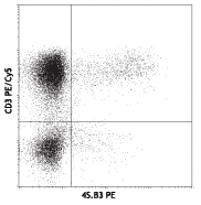
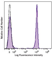
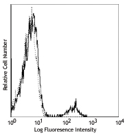
Compare Data Across All Formats
This data display is provided for general comparisons between formats.
Your actual data may vary due to variations in samples, target cells, instruments and their settings, staining conditions, and other factors.
If you need assistance with selecting the best format contact our expert technical support team.


Follow Us