 Login / Register


- Clone
- RPA-T8 (See other available formats)
- Regulatory Status
- RUO
- Workshop
- IV T171
- Other Names
- T8, Leu2
- Isotype
- Mouse IgG1, κ
- Ave. Rating
- Submit a Review
- Product Citations
- publications

-

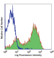
Human peripheral blood lymphocytes stained with RPA-T8 Alexa Fluor® 700
| Cat # | Size | Price | Quantity Check Availability | Save | ||
|---|---|---|---|---|---|---|
| 301027 | 25 µg | 126 CHF | ||||
| 301028 | 100 µg | 268 CHF | ||||
CD8a is a 32-34 kD type I glycoprotein. It forms a homodimer (CD8a/a) or heterodimer (CD8a/b) with CD8b. CD8, also known as T8 and Leu2, is a member of the immunoglobulin superfamily found on the majority of thymocytes, a subset of peripheral blood T cells, and NK cells (which express almost exclusively CD8a homodimers). CD8 acts as a co-receptor with MHC class I-restricted T cell receptors in antigen recognition and T cell activation, and has been shown to play a role in thymic differentiation. Two domains in CD8a are important for function: the extracellular IgSF domain binds the α3 domain of MHC class I and the cytoplasmic CXCP motif binds the tyrosine kinase p56 Lck.
Product DetailsProduct Details
- Reactivity
- Human,Cynomolgus,Rhesus
- Antibody Type
- Monoclonal
- Host Species
- Mouse
- Formulation
- Phosphate-buffered solution, pH 7.2, containing 0.09% sodium azide.
- Preparation
- The antibody was purified by affinity chromatography and conjugated with Alexa Fluor® 700 under optimal conditions.
- Concentration
- 0.5 mg/ml
- Storage & Handling
- The antibody solution should be stored undiluted between 2°C and 8°C, and protected from prolonged exposure to light. Do not freeze.
- Application
-
FC - Quality tested
- Recommended Usage
-
Each lot of this antibody is quality control tested by immunofluorescent staining with flow cytometric analysis. The suggested use of this reagent is ≤1.0 µg per million cells in 100 µl volume. It is highly recommended that the reagent be titrated for optimal performance for each application.
* Alexa Fluor® 700 has a maximum emission of 719 nm when it is excited at 633 nm / 635 nm. Prior to using Alexa Fluor® 700 conjugate for flow cytometric analysis, please verify your flow cytometer's capability of exciting and detecting the fluorochrome.
Alexa Fluor® and Pacific Blue™ are trademarks of Life Technologies Corporation.
View full statement regarding label licenses - Excitation Laser
-
Red Laser (633 nm)
- Application Notes
-
The RPA-T8 antibody does not block the binding of HIT8a antibody to CD8a. Additional reported applications of this antibody (for the relevant formats) include: immunohistochemical staining of paraformaldehyde-fixed frozen sections3 and costimulation of T cell responses4. This clone was tested in-house and does not work on formalin fixed paraffin-embedded (FFPE) tissue. The Ultra-LEAF™ purified antibody (Endotoxin <0.1 EU/µg, Azide-Free, 0.2 µm filtered) is recommended for functional assays (Cat. Nos. 301073 & 301074).
-
Application References
(PubMed link indicates BioLegend citation) -
- Knapp W, et al. Eds. 1989. Leucocyte Typing IV. Oxford University Press. New York.
- Schlossman S, et al. Eds. 1995. Leucocyte Typing V. Oxford University Press. New York.
- Mack CL, et al. 2004. Pediatr. Res. 56:79. (IHC)
- Magidovich E, et al. 2007. P. Natl. Acad. Sci. USA 104:13022.
- Thakarl D, et al. 2008.J. immunol. 180:7431. PubMed
- Kmieciak M, et al. 2009. J. Transl. Med. 7:89. (FC) PubMed
- Thakral D, et al. 2008. J. Immunol. 180:7431. (FC) PubMed
- Yoshino N, et al. 2000. Exp. Anim. (Tokyo) 49:97. (FC)
- Rout N, et al. 2010. PLoS One 5:e9787. (FC)
- Stoeckius M, et al. 2017. Nat. Methods. 14:865. (PG)
- Product Citations
- RRID
-
AB_493744 (BioLegend Cat. No. 301027)
AB_493745 (BioLegend Cat. No. 301028)
Antigen Details
- Structure
- Ig superfamily, homodimer or heterodimer with CD8β, 32-34 kD
- Distribution
-
Majority of thymocytes, T cell subset, NK cells
- Function
- MHC class I co-receptor, thymic differentiation, T cell activation
- Ligand/Receptor
- MHC Class I molecules
- Cell Type
- Dendritic cells, NK cells, T cells, Thymocytes, Tregs
- Biology Area
- Immunology
- Molecular Family
- CD Molecules
- Antigen References
-
1. Barclay N, et al. 1993. The Leucocyte Antigen FactsBook. Academic Press Inc. San Diego.
- Gene ID
- 925 View all products for this Gene ID
- UniProt
- View information about CD8alpha on UniProt.org
Related Pages & Pathways
Pathways


Customers Also Purchased
Compare Data Across All Formats
This data display is provided for general comparisons between formats.
Your actual data may vary due to variations in samples, target cells, instruments and their settings, staining conditions, and other factors.
If you need assistance with selecting the best format contact our expert technical support team.


Follow Us