 Login / Register


- Clone
- PC61 (See other available formats)
- Regulatory Status
- RUO
- Other Names
- IL-2Rα, Ly-43, p55, Tac
- Isotype
- Rat IgG1, λ
- Ave. Rating
- Submit a Review
- Product Citations
- publications

-

C57BL/6 mouse splenocytes were stained with CD4 FITC and CD25 (clone PC61) Brilliant Violet 650™.
| Cat # | Size | Price | Quantity Check Availability | Save | ||
|---|---|---|---|---|---|---|
| 102037 | 125 µL | 226 CHF | ||||
| 102038 | 50 µg | 273 CHF | ||||
CD25 is a 55 kD glycoprotein also known as the low affinity IL-2Rα, Ly-43, p55, or Tac. It is expressed on activated T and B cells, thymocyte subsets, pre-B cells, and T regulatory cells. In association with CD122 (IL-2Rβ) and CD132 (common γ chain), CD25 forms the high affinity signaling IL-2 receptor.
Product DetailsProduct Details
- Reactivity
- Mouse
- Antibody Type
- Monoclonal
- Host Species
- Rat
- Immunogen
- IL-2-dependent cytolytic mouse T-cell clone B6.1
- Formulation
- Phosphate-buffered solution, pH 7.2, containing 0.09% sodium azide and BSA (origin USA).
- Preparation
- The antibody was purified by affinity chromatography and conjugated with Brilliant Violet 650™ under optimal conditions.
- Concentration
- µg sizes: 0.2 mg/mLµL sizes: lot-specific (to obtain lot-specific concentration and expiration, please enter the lot number in our Certificate of Analysis online tool.)
- Storage & Handling
- The antibody solution should be stored undiluted between 2°C and 8°C, and protected from prolonged exposure to light. Do not freeze.
- Application
-
FC - Quality tested
- Recommended Usage
-
Each lot of this antibody is quality control tested by immunofluorescent staining with flow cytometric analysis. For immunofluorescent staining using the µg size, the suggested use of this reagent is ≤0.25 µg per million cells in 100 µl volume. For immunofluorescent staining using the µl size, the suggested use of this reagent is 5 µl per million cells in 100 µl staining volume or 5 µl per 100 µl of whole blood. It is recommended that the reagent be titrated for optimal performance for each application.
Brilliant Violet 650™ excites at 405 nm and emits at 645 nm. The bandpass filter 660/20 nm is recommended for detection, although filter optimization may be required depending on other fluorophores used. Be sure to verify that your cytometer configuration and software setup are appropriate for detecting this channel. Refer to your instrument manual or manufacturer for support. Brilliant Violet 650™ is a trademark of Sirigen Group Ltd.
Learn more about Brilliant Violet™.
This product is subject to proprietary rights of Sirigen Inc. and is made and sold under license from Sirigen Inc. The purchase of this product conveys to the buyer a non-transferable right to use the purchased product for research purposes only. This product may not be resold or incorporated in any manner into another product for resale. Any use for therapeutics or diagnostics is strictly prohibited. This product is covered by U.S. Patent(s), pending patent applications and foreign equivalents. - Excitation Laser
-
Violet Laser (405 nm)
- Application Notes
-
Additional reported applications (for the relevant formats) include: immunoprecipitation1,2, in vitro blocking of IL-2 binding to low- and high-affinity receptors1-4, growth inhibition of IL-2-dependent T-cell lines1-4, in vivo depletion of CD25+CD4+ Treg cells5-8,10, and immunohistochemical staining of acetone-fixed frozen sections2. PC61 antibody recognizes a different epitope than 3C7 antibody (Cat. No. 101902). For in vivo studies or highly sensitive assays, we recommend Ultra-LEAF™ purified antibody (Cat. No. 102040) with endotoxin < 0.01 EU/µg, Azide-Free, 0.2 µm filtered.
-
Application References
(PubMed link indicates BioLegend citation) -
- Lowenthal JW, et al. 1985. Nature 315:669. (IP, Block)
- Ceredig R, et al. 1985. Nature 314:98. (IP, IHC, Block)
- Lowenthal JW, et al. 1985. J. Immunol. 135:3988. (Block)
- Moreau JL, et al. 1987. Eur. J. Immunol. 17:929. (Block)
- Takahashi T, et al. 2000. J. Exp. Med. 192:303. (Deplete)
- Onizuka S, et al. 1999. Cancer Res. 59:3128. (Deplete)
- Lei TC, et al. 2005. Blood 105:4865. (Deplete)
- Pasare C, et al. 2004. Immunity 21:733. (Deplete)
- León-Ponte M, et al. 2007. Blood 109:3139.
- Cao OW, et al. 2007. Blood doi:10.1182/blood-2007-02-073304. (Deplete)
- Benson MJ, et al. 2007. J. Exp. Med. doi:10.1084/jem.20070719.
- Liu F, et al. 2011. Arch Toxicol. 85:1383. PubMed
- Anguela XM, et al. 2013. Diabetes. 62:551. PubMed
- Product Citations
- RRID
-
AB_11125760 (BioLegend Cat. No. 102037)
AB_2563060 (BioLegend Cat. No. 102038)
Antigen Details
- Structure
- Forms high affinity IL-2R with IL-2Rβ (CD122) and IL-2Rγ (CD132), 55 kD
- Distribution
-
Activated T cells and B cells, thymocyte subset, pre-B cells, T regulatory cells
- Function
- IL-2 receptor
- Ligand/Receptor
- IL-2
- Cell Type
- B cells, T cells, Thymocytes, Tregs
- Biology Area
- Immunology
- Molecular Family
- CD Molecules, Cytokine/Chemokine Receptors
- Antigen References
-
- Taniguchi T, et al. 1993. Cell 73:5-8.
- Waldmann TA. 1991. J Biol Chem. 266:2681-4.
- Read S, et al. 2000. J Exp Med. 192:295-302.
- Lowenthal JW, et al. 1985. J Immunol. 135:3988-94.
- Gene ID
- 16184 View all products for this Gene ID
- UniProt
- View information about CD25 on UniProt.org
Related Pages & Pathways
Pathways


Customers Also Purchased
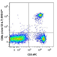
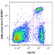
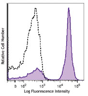
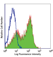
Compare Data Across All Formats
This data display is provided for general comparisons between formats.
Your actual data may vary due to variations in samples, target cells, instruments and their settings, staining conditions, and other factors.
If you need assistance with selecting the best format contact our expert technical support team.


Follow Us