 Login / Register


- Clone
- 30-F11 (See other available formats)
- Regulatory Status
- RUO
- Other Names
- T200, Ly-5, LCA
- Isotype
- Rat IgG2b, κ
- Ave. Rating
- Submit a Review
- Product Citations
- publications

-

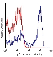
C57BL/6 mouse splenocytes were stained with CD45 (clone 30-F11) Brilliant Violet 785™ (filled histogram) or rat IgG2b, κ Brilliant Violet 785™ isotype control (open histogram).
| Cat # | Size | Price | Quantity Check Availability | Save | ||
|---|---|---|---|---|---|---|
| 103149 | 50 µg | 273 CHF | ||||
CD45 is a 180-240 kD glycoprotein also known as the leukocyte common antigen (LCA), T200, or Ly-5. It is a member of the protein tyrosine phosphatase (PTP) family, expressed on all hematopoietic cells except mature erythrocytes and platelets. There are different isoforms of CD45 that arise from variable splicing of exons 4, 5, and 6, which encode A, B, and C determinants, respectively. CD45 plays a key role in TCR and BCR signal transduction. These isoforms are very specific to the activation and maturation state of the cell as well as cell type. The primary ligands for CD45 are galectin-1, CD2, CD3, CD4, TCR, CD22, and Thy-1.
Product DetailsProduct Details
- Reactivity
- Mouse
- Antibody Type
- Monoclonal
- Host Species
- Rat
- Immunogen
- Mouse thymus or spleen
- Formulation
- Phosphate-buffered solution, pH 7.2, containing 0.09% sodium azide and BSA (origin USA).
- Preparation
- The antibody was purified by affinity chromatography and conjugated with Brilliant Violet 785™ under optimal conditions.
- Concentration
- 0.2 mg/ml
- Storage & Handling
- The antibody solution should be stored undiluted between 2°C and 8°C, and protected from prolonged exposure to light. Do not freeze.
- Application
-
FC - Quality tested
- Recommended Usage
-
Each lot of this antibody is quality control tested by immunofluorescent staining with flow cytometric analysis. For flow cytometric staining, the suggested use of this reagent is ≤0.5 µg per million cells in 100 µl volume. It is recommended that the reagent be titrated for optimal performance for each application.
Brilliant Violet 785™ excites at 405 nm and emits at 785 nm. The bandpass filter 780/60 nm is recommended for detection, although filter optimization may be required depending on other fluorophores used. Be sure to verify that your cytometer configuration and software setup are appropriate for detecting this channel. Refer to your instrument manual or manufacturer for support. Brilliant Violet 785™ is a trademark of Sirigen Group Ltd.
Learn more about Brilliant Violet™.
This product is subject to proprietary rights of Sirigen Inc. and is made and sold under license from Sirigen Inc. The purchase of this product conveys to the buyer a non-transferable right to use the purchased product for research purposes only. This product may not be resold or incorporated in any manner into another product for resale. Any use for therapeutics or diagnostics is strictly prohibited. This product is covered by U.S. Patent(s), pending patent applications and foreign equivalents. - Excitation Laser
-
Violet Laser (405 nm)
- Application Notes
-
Clone 30-F11 reacts with all isoforms and both CD45.1 and CD45.2 alloantigens of CD45.
Additional reported applications (for relevant formats) include: immunoprecipitation3, complement-dependent cytotoxicity1,5, immunohistochemistry (acetone-fixed frozen sections, zinc-fixed paraffin-embedded sections and formalin-fixed paraffin-embedded sections)4,6, Western blotting7, and spatial biology (IBEX)10,11. The Ultra-LEAF™ purified antibody (Endotoxin < 0.01 EU/µg, Azide-Free, 0.2 µm filtered) is recommended for functional assays (Cat. No. 103163 and 103164). -
Application References
(PubMed link indicates BioLegend citation) -
- Podd BS, et al. 2006. J. Immunol. 176:6532. (FC, CMCD) PubMed
- Haynes NM, et al. 2007. J. Immunol. 179:5099. (FC)
- Ledbetter JA, et al. 1979. Immunol. Rev. 47:63. (IP)
- Simon DI, et al. 2000. J. Clin. Invest. 105:293. (IHC)
- Seaman WE. 1983. J. Immunol. 130:1713. (CMCD)
- Cornet A, et al. 2001. P. Natl. Acad. Sci. USA 98:13306. (IHC)
- Tsuboi S and Fukuda M. 1998. J. Biol. Chem. 273:30680. (WB) PubMed
- Liu F, et al. 2012. Blood. 119:3295. PubMed
- Pelletier AN, et al. 2012. J. Immunol. 188:5561. PubMed
- Radtke AJ, et al. 2020. Proc Natl Acad Sci U S A. 117:33455-65. (SB) PubMed
- Radtke AJ, et al. 2022. Nat Protoc. 17:378-401. (SB) PubMed
- Product Citations
- RRID
-
AB_2564590 (BioLegend Cat. No. 103149)
Antigen Details
- Structure
- Protein tyrosine phosphatase (PTP) family, 180-240 kD
- Distribution
-
All hematopoietic cells except mature erythrocytes and platelets
- Function
- Phosphatase, T and B cell activation
- Ligand/Receptor
- Galectin-1, CD2, CD3, CD4, TCR, CD22, Thy-1
- Cell Type
- B cells, Dendritic cells, Mesenchymal Stem Cells, Tregs
- Biology Area
- Cell Biology, Immunology, Inhibitory Molecules, Innate Immunity, Neuroscience, Neuroscience Cell Markers, Stem Cells
- Molecular Family
- CD Molecules
- Antigen References
-
1. Barclay A, et al. 1997. The Leukocyte Antigen FactsBook Academic Press.
2. Trowbridge IS, et al. 1993. Annu. Rev. Immunol. 12:85.
3. Kishihara K, et al. 1993. Cell 74:143.
4. Pulido R, et al. 1988. J. Immunol. 140:3851. - Gene ID
- 19264 View all products for this Gene ID
- UniProt
- View information about CD45 on UniProt.org
Related Pages & Pathways
Pathways


Customers Also Purchased
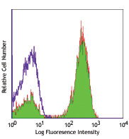
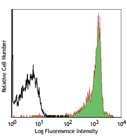
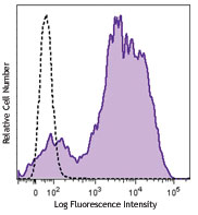
Compare Data Across All Formats
This data display is provided for general comparisons between formats.
Your actual data may vary due to variations in samples, target cells, instruments and their settings, staining conditions, and other factors.
If you need assistance with selecting the best format contact our expert technical support team.


Follow Us