 Login / Register


- Clone
- 22F6 (See other available formats)
- Regulatory Status
- RUO
- Other Names
- IKZF2, IKAROS family zinc finger 2, ANF1A2, ZNF1A2, ZNFN1A2
- Isotype
- Armenian Hamster IgG
- Ave. Rating
- Submit a Review
- Product Citations
- publications

-

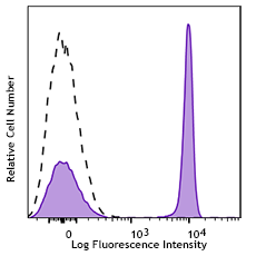
C57BL/6 splenocytes surface stained with CD4-APC (GK1.5), and then intracellularly stained with Helios-PE (clone 22F6). -

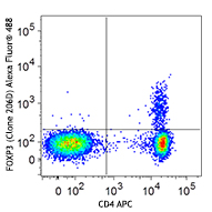
Human peripheral blood lymphocytes surface stained with CD4-APC (RPA-T4), and then intracellulary stained with Helios-PE (clone 22F6).
| Cat # | Size | Price | Quantity Check Availability | Save | ||
|---|---|---|---|---|---|---|
| 137206 | 25 tests | 214 CHF | ||||
| 137216 | 100 tests | 420 CHF | ||||
Helios is a member of the Ikaros family of zinc finger transcription factors. It contains a C-terminal region composed of 2 zinc-finger domains that mediate dimerization between the family members. Helios was originally cloned from a mouse thymoma line. It is mainly expressed in peripheral T cells and thymocytes. It is found at high levels in a subpopulation of regulatory T cells. Helios plays an important role in T cell development and homeostasis. Overexpression of Helios profoundly alters αβ T cell differentiation and activation. It has been determined that silencing of Helios in B cells is critical for maintaining normal B cell function. Helios is also involved in tumor immunity.
Product DetailsProduct Details
- Reactivity
- Mouse,Human
- Antibody Type
- Monoclonal
- Host Species
- Armenian Hamster
- Immunogen
- Helios peptide (aa 51-107)
- Formulation
- Phosphate-buffered solution, pH 7.2, containing 0.09% sodium azide and BSA (origin USA)
- Preparation
- The antibody was purified by affinity chromatography, and conjugated with PE under optimal conditions.
- Concentration
- Lot-specific (to obtain lot-specific concentration and expiration, please enter the lot number in our Certificate of Analysis online tool.)
- Storage & Handling
- The antibody solution should be stored undiluted between 2°C and 8°C, and protected from prolonged exposure to light. Do not freeze.
- Application
-
ICFC - Quality tested
- Recommended Usage
-
Each lot of this antibody is quality control tested by intracellular flow cytometry using our True-Nuclear™ Transcription Factor Staining Protocol. It is recommended that the reagent be titrated for optimal performance for each application.
For flow cytometric staining, the suggested use of this reagent is 5 µl per million cells in 100 µl staining volume or 5 µl per 100 µl of whole blood. - Excitation Laser
-
Blue Laser (488 nm)
Green Laser (532 nm)/Yellow-Green Laser (561 nm)
- Application Notes
-
NOTE: For flow cytometric staining with this clone, True-Nuclear™ Transcription Factor Buffer Set (Cat. No. 424401) offers improved staining and is highly recommended over the Foxp3 Fix/Perm Buffer Set (Cat. No. 421403) and the True-Nuclear™ 10X Perm Buffer (Cat. No. 424403).
View more applications data for a different format of this clone in our Scientific Poster Library. -
Application References
(PubMed link indicates BioLegend citation) -
- Thornton AM, et al. 2010. J. Immunol. 184:1. PubMed
- Verhagen J and Wraith D. 2010. J. Immunol. 185:7129.
- Stone B, et al. 2012. Clin Immunol. 145:153. PubMed
- Vaeth M, et al. 2012. PNAS. 109:16258. PubMed
- Angin M, et al. 2014. PLoS One. 9:86920. PubMed
- Bedke T, et al. 2014. Immunol Cell Biol. PubMed
- Liu Y, et al. 2014. Am J Physiol Gastrointest Liver Physiol. 307:177. PubMed
- Verhagen J and Wraith DC. 2014. J. Immunol. Methods. S0022. (FC) PubMed
- Product Citations
- RRID
-
AB_10552903 (BioLegend Cat. No. 137206)
AB_10660749 (BioLegend Cat. No. 137216)
Antigen Details
- Structure
- A zinc finger nuclear transcription factor of the Ikaros family, containing 2 zinc-finger domains in C-terminal region.
- Distribution
-
Regulatory T cells (Tregs), conventional T cells, thymocytes.
- Function
- Plays an important role in T cell development and homeostasis, involved in tumor immunity.
- Cell Type
- T cells, Thymocytes, Tregs
- Biology Area
- Cell Biology, Immunology, Transcription Factors
- Molecular Family
- Nuclear Markers
- Antigen References
-
1. Kelly CM, et al. 1998. Curr. Biol. 8:508.
2. Dovat S, et al. 2005. J. Immunol. 175:3508.
3. Cortes M, et al. 1999. Curr. Opin. Immunol. 11:167.
4. Cai Q, et al. 2009. J. Immunol. 183:2303.
5. Zhang Z, et al. 2007. Blood 109:2190.
6. Hahm K, et al. 1998. Genes Dev. 12:782. - Gene ID
- 22779 View all products for this Gene ID 22807 View all products for this Gene ID
- UniProt
- View information about Helios on UniProt.org
Related Pages & Pathways
Pathways


Related FAQs
- What type of PE do you use in your conjugates?
- We use R-PE in our conjugates.
Customers Also Purchased
Compare Data Across All Formats
This data display is provided for general comparisons between formats.
Your actual data may vary due to variations in samples, target cells, instruments and their settings, staining conditions, and other factors.
If you need assistance with selecting the best format contact our expert technical support team.


Follow Us