 Login / Register


- Clone
- G025H7 (See other available formats)
- Regulatory Status
- RUO
- Other Names
- CXCR3, G protein-coupled receptor 9 (GPR9), CKR-L2
- Isotype
- Mouse IgG1, κ
- Ave. Rating
- Submit a Review
- Product Citations
- publications

-

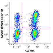
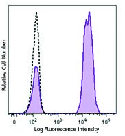
Human peripheral lymphocytes were stained with CD3 FITC and CXCR3 (clone G025H7) PE/Cyanine7 (left) or mouse IgG1, κ PE/Cyanine7 isotype control (right).
| Cat # | Size | Price | Quantity Check Availability | Save | ||
|---|---|---|---|---|---|---|
| 353719 | 25 tests | 159 CHF | ||||
| 353720 | 100 tests | 393 CHF | ||||
Human CXCR3, also known as GPR9, is a chemokine receptor that binds CXCL9, CXCL10, and CXCL11. It is a 38 kD seven-pass transmembrane receptor coupled to G-protein. CXCR3 is highly expressed by T cells (Th1), natural killer cells (NK cells), dendritic cells, mast cells, alveolar macrophages, eosinophils, and human airway epithelial cells. CXCR3 is important for effector lymphocyte recruitment into inflamed tissue in various inflammatory and autoimmune diseases, such as chronically inflamed liver, Crohn's disease, rheumatoid arthritis, multiple sclerosis, and inflammatory skin diseases.
Product DetailsProduct Details
- Reactivity
- Human,Cynomolgus,Rhesus
- Antibody Type
- Monoclonal
- Host Species
- Mouse
- Immunogen
- Human CXCR3 transfectants
- Formulation
- Phosphate-buffered solution, pH 7.2, containing 0.09% sodium azide and BSA (origin USA)
- Preparation
- The antibody was purified by affinity chromatography and conjugated with PE/Cyanine7 under optimal conditions.
- Concentration
- Lot-specific (to obtain lot-specific concentration and expiration, please enter the lot number in our Certificate of Analysis online tool.)
- Storage & Handling
- The antibody solution should be stored undiluted between 2°C and 8°C, and protected from prolonged exposure to light. Do not freeze.
- Application
-
FC - Quality tested
- Recommended Usage
-
Each lot of this antibody is quality control tested by immunofluorescent staining with flow cytometric analysis. For flow cytometric staining, the suggested use of this reagent is 5 µl per million cells in 100 µl staining volume or 5 µl per 100 µl of whole blood.
- Excitation Laser
-
Blue Laser (488 nm)
Green Laser (532 nm)/Yellow-Green Laser (561 nm)
- Additional Product Notes
-
BioLegend is in the process of converting the name PE/Cy7 to PE/Cyanine7. The dye molecule remains the same, so you should expect the same quality and performance from our PE/Cyanine7 products. Please contact Technical Service if you have any questions.
- Product Citations
- RRID
-
AB_11218804 (BioLegend Cat. No. 353719)
AB_11219383 (BioLegend Cat. No. 353720)
Antigen Details
- Structure
- CXC-chemokine receptor, G protein-coupled receptor, seven-pass transmembrane receptor
- Distribution
-
T cell subset, NK cells, plasmacytoid dendritic cells, GM-CSF activated CD34+ hematopoietic progenitors, mast cells, alveolar macrophages, eosinophils, and airway epithelial cells
- Function
- Essential in T cell recruitment to sites of inflammation
- Ligand/Receptor
- CXCL9, CXCL10, and CXCL11
- Cell Type
- Dendritic cells, Eosinophils, Epithelial cells, Hematopoietic stem and progenitors, Macrophages, Mast cells, NK cells, T cells, Tregs
- Biology Area
- Cell Biology, Immunology, Neuroinflammation, Neuroscience
- Molecular Family
- CD Molecules, Cytokine/Chemokine Receptors, GPCR
- Antigen References
-
1. Loetscher M, et al. 1996. J. Exp. Med. 184:963.
2. Cole KE, et al. 1998. J. Exp. Med. 187:2009.
3. Aksoy MO, et al. 2006. Am. J. Physiol. Lung Cell Mol. Physiol. 290:L909.
4. Curbishley SM, et al. 2005. Am. J. Pathol. 167:887.
5. Turner JE, et al. 2007. Mini. Rev. Med. Chem. 7:1089.
6. Wenzel J, et al. 2008. J. Invest. Dermatol. 128:67. - Gene ID
- 2833 View all products for this Gene ID
- UniProt
- View information about CD183 on UniProt.org
Related Pages & Pathways
Pathways


Related FAQs
- Does staining at room temperature or even at 37°C help for checking chemokine receptors expression?
-
Due to continuous recycling of many chemokine receptors, it may be worthwhile to consider staining at room temperature or at 37°C if the staining at lower temperature (which can potentially reduce receptor turnover) is not optimal.
Customers Also Purchased
Compare Data Across All Formats
This data display is provided for general comparisons between formats.
Your actual data may vary due to variations in samples, target cells, instruments and their settings, staining conditions, and other factors.
If you need assistance with selecting the best format contact our expert technical support team.


Follow Us