 Login / Register


- Clone
- L5 (See other available formats)
- Regulatory Status
- RUO
- Other Names
- FLAG tag
- Isotype
- Rat IgG2a, λ
- Ave. Rating
- Submit a Review
- Product Citations
- publications

-

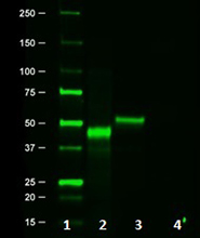
Cell extracts from untransfected 293T cells (lane 1) or 293T cells transfected with a plasmid encoding DYKDDDDK-tagged protein (lane 2), using anti-DYKDDDDK, clone L5.
The DYKDDDDK tag, commonly referred to as Sigma®'s FLAG® Tag, is often used as a protein modification in order to simplify the labeling and detection of proteins.This unique amino acid sequence allows for specific antibody detection in western blotting, immunoprecipitation, and immunostaining techniques.Due to the short sequence, this modification is not likely to affect the structure or function of the modified proteins.
Product DetailsProduct Details
- Reactivity
- Epitope tag
- Antibody Type
- Monoclonal
- Host Species
- Rat
- Immunogen
- DYKDDDDK-tagged mouse Langerin
- Formulation
- Phosphate-buffered solution, pH 7.2, containing 0.09% sodium azide.
- Preparation
- The antibody was purified by affinity chromatography.
- Concentration
- 0.5 mg/ml
- Storage & Handling
- The antibody solution should be stored undiluted between 2°C and 8°C.
- Application
-
WB - Quality tested
IP, ICC, ELISA, FC, Purification - Reported in the literature, not verified in house - Recommended Usage
-
Each lot of this antibody is quality control tested by Western blotting. For Western blotting, suggested working dilution(s): Use 5 µg per 5 ml antibody dilution buffer for each mini-gel. It is recommended that the reagent be titrated for optimal performance for each application.
- Application Notes
-
The L5 clone has been demonstrated to have 2-8 fold better sensitivity in WB than another commonly used antibody clone, M2.
-
Application References
(PubMed link indicates BioLegend citation) -
- Park SH, et al. 2008. J Immunol Methods. 331:27.
- Moon SH, et al. 2010. J. Biol Chem. 285:12935. PubMed
- Sasaki M, et al. 2011. J. Biol Chem. 286:39370. PubMed
- Sonder SU, et al. 2012. J Immunol. 188:5906. PubMed
- Jiang Y, et al. 2013. Int Immunol. 25:235. PubMed
- Zuo X, et al. 2014. PLoS One. 9:84748. PubMed
- Toyo-Oak K, et al. 2014. J Neurosci. 34:12168. PubMed
- Product Citations
- RRID
-
AB_2749907 (BioLegend Cat. No. 637319)
AB_1134266 (BioLegend Cat. No. 637301)
AB_1134268 (BioLegend Cat. No. 637302)
AB_1134265 (BioLegend Cat. No. 637303)
AB_1134267 (BioLegend Cat. No. 637304)
Antigen Details
- Biology Area
- Cell Biology
- Antigen References
-
1. Einhauer A. 2001. J. Biochem. Biophys. Methods. 49:455.
2. Knappik A and Pluckthun A. 1994. Biotechniques. 17:754. - Gene ID
- NA
- UniProt
- View information about DYKDDDDK on UniProt.org

Related FAQs
- I am unable to detect FLAG tag with L5 antibody clone by IP and/or western blotting.
-
Some points to consider here include
- Sometimes changing tag location (form N-terminus to C-terminus or vice versa) or use tandem FLAG tag (3x) can also increase the chances of tag detection
- Tagged protein is either not expressed or lowly expressed
- Always include a positive control to make sure the process of working fine
- Does anti tag antibody recognize the tag sequence at N terminal or C terminal or within the fusion protein?
- Our antibody can recognize the fusion protein with tag either at N-terminus or C-terminus or within the protein if the tag is exposed.
Customers Also Purchased
Compare Data Across All Formats
This data display is provided for general comparisons between formats.
Your actual data may vary due to variations in samples, target cells, instruments and their settings, staining conditions, and other factors.
If you need assistance with selecting the best format contact our expert technical support team.


Follow Us