 Login / Register


- Clone
- W17079A (See other available formats)
- Regulatory Status
- RUO
- Other Names
- glyceraldehyde-3-phosphate dehydrogenase
- Isotype
- Rat IgG2a, κ
- Ave. Rating
- Submit a Review
- Product Citations
- publications

-

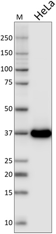
Whole cell lysates (15 µg protein) from K562, PC3, HeLa, Molt-4, NTRA-2, NIH3T3, and UMR-106 cells were resolved by electrophoresis (4-12% Bis-Tris gel), transferred to nitrocellulose, and probed with 1:10000 (0.05 µg/mL) Purified anti-GAPDH Antibody, clone W17079A. Proteins were visualized using chemiluminescence detection by incubating with 1:3000 dilution of HRP goat anti-rat-IgG secondary antibody (Cat. No. 405405, upper). 1:2000 dilution of Direct-Blot™ HRP anti-β-actin antibody (Cat. No. 643807) was used as a loading control (lower). Lane M: MW ladder. -

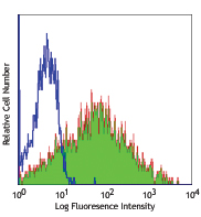
HeLa cells were fixed with cold methanol for 15 minutes and blocked with 5% FBS for 60 minutes. Then the cells were intracellularly stained with (A) 1:500 (1 µg/ml) purified rat IgG2a, κ isotype control antibody (Cat. No. 400502) or 1:500 (1 µg/ml, B), 1:2000 (0.25 µg/ml, C) and 1:5000 (0.1 µg/ml, D) anti-GAPDH antibody (clone W17079A) overnight at 4°C followed by Alexa Fluor® 488 (green) conjugated goat anti-rat IgG (Cat. No. 405418) for one hour at room temperature. Nuclei were counterstained with DAPI (blue). The image was captured with a 60X objective.
| Cat # | Size | Price | Quantity Check Availability | Save | ||
|---|---|---|---|---|---|---|
| 607901 | 25 µg | 101 CHF | ||||
| 607902 | 100 µg | 253 CHF | ||||
GAPDH is well known for its glycolytic function of converting D-glyceraldehyde-3-phosphate to 1,3-bisphosphoglycerate. GAPDH is a ubiquitously expressed and has a molecular mass of 36 kD. Though differentially expressed from tissue to tissue, GAPDH is frequently used as a loading control for assays involving mRNA and protein detection. In more recent studies, GAPDH has been shown to be involved in microtubule bundling, prostate cancer progression, programmed neuronal cell death, DNA replication, and DNA repair. Recent work has elucidated roles for GAPDH in apoptosis, gene expression and nuclear transport. GAPDH may also play a role in neurodegenerative pathologies such as Huntington and Alzheimer's diseases.
Product DetailsProduct Details
- Reactivity
- Human,Mouse
- Antibody Type
- Monoclonal
- Host Species
- Rat
- Immunogen
- Recombinant protein (161-335 a.a.)
- Formulation
- Phosphate-buffered solution, pH 7.2, containing 0.09% sodium azide.
- Preparation
- The antibody was purified by affinity chromatography.
- Concentration
- 0.5 mg/ml
- Storage & Handling
- The antibody solution should be stored undiluted between 2°C and 8°C.
- Application
-
WB - Quality tested
ICC - Verified - Recommended Usage
-
Each lot of this antibody is quality control tested by Western blotting. For Western blotting, the suggested use of this reagent is 0.0375-0.5 µg per ml (1:1000-1:16000 dilution). For immunocytochemistry, a concentration range of 0.25-1.0 μg/ml (1:500-1:2000 dilution) is recommended. It is recommended that the reagent be titrated for optimal performance for each application.
- Application Notes
-
This clone weakly reacts with rat GAPDH.
- Product Citations
- RRID
-
AB_2734502 (BioLegend Cat. No. 607901)
AB_2734503 (BioLegend Cat. No. 607902)
Antigen Details
- Structure
- Belongs to the glyceraldehyde-3-phosphate dehydrogenase family, predicted molecular weight 36 kD. The enzyme exists as a tetramer of identical chains.
- Distribution
-
Ubiquitously expressed, locate in cytoplasm and perinuclear region.
- Function
- Belongs to the glyceraldehyde-3-phosphate dehydrogenase family, predicted molecular weight 36 kD. The enzyme exists as a tetramer of identical chains.
- Interaction
- Interacts with TPPP. Interacts with EIF1AD and WARS. Interacts with SUMO4, GLUT4, nPKC-iota and CAMK2.
- Biology Area
- Cell Biology, Western Blot Controls
- Antigen References
-
- Ercolani L, et al. 1988. J. Biol. Chem. 263:15335.
- Meyer-Siegler K, et al. 1991. Proc. Natl. Acad. Sci. USA. 88:8460.
- Hara MR, Snyder SH. 2006. Cell. Mol. Neurobiol. 26:527.
- Zheng L, et al. 2003. Cell. 114:255.
- Wang Q, et al. 2005. FASEB J. 19:869.
- Bae BI, et al. 2006. Proc. Natl. Acad. Sci. USA. 103:3405.
- Gene ID
- 5590 View all products for this Gene ID
- UniProt
- View information about GAPDH on UniProt.org

Related FAQs
Customers Also Purchased
Compare Data Across All Formats
This data display is provided for general comparisons between formats.
Your actual data may vary due to variations in samples, target cells, instruments and their settings, staining conditions, and other factors.
If you need assistance with selecting the best format contact our expert technical support team.


Follow Us