 Login / Register


- Clone
- A1 (See other available formats)
- Regulatory Status
- RUO
- Workshop
- HCDM listed
- Other Names
- gp80, E-ATPDase, NTPDase-1, ecto-apyrase, Ec3.6.1.5
- Isotype
- Mouse IgG1, κ
- Ave. Rating
- Submit a Review
- Product Citations
- publications

-

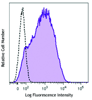
Human peripheral blood lymphocytes stained with purified A1, followed by anti-mouse IgG FITC
| Cat # | Size | Price | Quantity Check Availability | Save | ||
|---|---|---|---|---|---|---|
| 328202 | 100 µg | 112 CHF | ||||
Human CD39 is an integral membrane protein with two transmembrane domains. It exists as a homotetramer. Expression of CD39 is found on activated lymphocytes, a subset of T cells and B cells, and dendritic cells with weak staining on monocytes and granulocytes. CD39 and CD73 have been found on regulatory T cells, specifically the effector/memory like T cells. CD39 can hydrolyze both nucleoside triphosphates and diphosphates. CD39 is the dominant ecto nucleotidase of vascular and placental trophoblastic tissues and appears to modulate the functional expression of type 2 purinergic (P2) G protein coupled receptors (GPCRs). CD39 has intrinsic ecto-ATPase activity. Expression of CD39 is induced on T cells and increased on B cells as a late activation antigen.
Product DetailsProduct Details
- Reactivity
- Human,Cynomolgus,Rhesus
- Antibody Type
- Monoclonal
- Host Species
- Mouse
- Immunogen
- PHA activated human lymphocytes
- Formulation
- Phosphate-buffered solution, pH 7.2, containing 0.09% sodium azide.
- Preparation
- The antibody was purified by affinity chromatography.
- Concentration
- 0.5 mg/ml
- Storage & Handling
- The antibody solution should be stored undiluted between 2°C and 8°C.
- Application
-
FC - Quality tested
IHC-P, Block - Reported in the literature, not verified in house - Recommended Usage
-
Each lot of this antibody is quality control tested by immunofluorescent staining with flow cytometric analysis. For flow cytometric staining, the suggested use of this reagent is ≤ 1.0 µg per 106 cells in 100 µl volume or 100 µl of whole blood. It is recommended that the reagent be titrated for optimal performance for each application.
- Application Notes
-
The A1 antibody binds to the human CD39 cell surface antigen and has been shown to block MHC independent target cell recognition by hapten-specific CTL. Additional reported applications (for the relevant formats) include: in vitro CD39 blockade3, immunofluorescence4, immunohistochemistry6, and spatial biology (IBEX)7,8. The Ultra-LEAF™ purified antibody (Endotoxin < 0.01 EU/µg, Azide-Free, 0.2 µm filtered) is recommended for blocking assays (contact our custom solutions team).
-
Application References
(PubMed link indicates BioLegend citation) -
- Aversa GG, et al. 1988. Transplant. P. 20:4952.
- Aversa GG, et al. 1989. Transplant. P. 21:34950.
- Borsellino G, et al. 2007. Blood. 110:1225. (Block)
- Stockl J, et al. 2001. J. Immunol. 167:2724. (IF)
- Sestak K, et al. 2007. Vet. Immunol. Immunopathol. 119:21.
- Lyck L, et al. 2008. J. Histochem. Cytochem. 56:201. (IHC)
- Radtke AJ, et al. 2020. Proc Natl Acad Sci USA. 117:33455-33465. (SB) PubMed
- Radtke AJ, et al. 2022. Nat Protoc. 17:378-401. (SB) PubMed
- Product Citations
- RRID
-
AB_940438 (BioLegend Cat. No. 328202)
Antigen Details
- Distribution
-
Activated lymphocytes, and also on a subset of T cells, regulatory T cells, B cells, and dendritic cells.
- Cell Type
- B cells, Dendritic cells, Lymphocytes, T cells, Tregs
- Biology Area
- Immunology
- Molecular Family
- CD Molecules
- Gene ID
- 953 View all products for this Gene ID
- UniProt
- View information about CD39 on UniProt.org


Customers Also Purchased
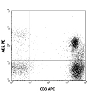
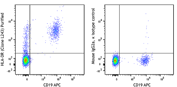
Compare Data Across All Formats
This data display is provided for general comparisons between formats.
Your actual data may vary due to variations in samples, target cells, instruments and their settings, staining conditions, and other factors.
If you need assistance with selecting the best format contact our expert technical support team.


Follow Us