 Login / Register


- Clone
- 5E10 (See other available formats)
- Regulatory Status
- RUO
- Workshop
- HCDM listed
- Other Names
- Thy-1, Thy1
- Isotype
- Mouse IgG1, κ
- Ave. Rating
- Submit a Review
- Product Citations
- publications

-

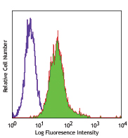
Human erythroleukemic cell line (HEL) stained with 5E10 FITC
| Cat # | Size | Price | Quantity Check Availability | Save | ||
|---|---|---|---|---|---|---|
| 328107 | 25 tests | 81€ | ||||
| 328108 | 100 tests | 184€ | ||||
CD90 is a 25-35 kD GPI-anchored protein, also known as Thy-1. It belongs to the Ig superfamily. Human CD90 is expressed on neuronal cells, a subset of CD34+ cells, a subset of fetal liver cells and fetal thymocytes, fibroblasts, activated endothelial cells, and some leukemia cell lines. CD34+CD90+ cells are primitive hematopoietic stem cells. It has been reported that Thy-1 binds with β2 and β3 integrins and plays bimodal roles in the regulation of cell adhesion and neurite outgrowth, and inhibits hematopoietic stem cells proliferation and differentiation.
Product DetailsProduct Details
- Verified Reactivity
- Human
- Reported Reactivity
- African Green, Baboon, Cynomolgus, Pigtailed Macaque, Rhesus, Pig
- Antibody Type
- Monoclonal
- Host Species
- Mouse
- Immunogen
- HEL cells
- Formulation
- Phosphate-buffered solution, pH 7.2, containing 0.09% sodium azide and BSA (origin USA)
- Preparation
- The antibody was purified by affinity chromatography, and conjugated with FITC under optimal conditions.
- Concentration
- Lot-specific (to obtain lot-specific concentration and expiration, please enter the lot number in our Certificate of Analysis online tool.)
- Storage & Handling
- The antibody solution should be stored undiluted between 2°C and 8°C, and protected from prolonged exposure to light. Do not freeze.
- Application
-
FC - Quality tested
- Recommended Usage
-
Each lot of this antibody is quality control tested by immunofluorescent staining with flow cytometric analysis. For flow cytometric staining, the suggested use of this reagent is 5 µl per million cells in 100 µl staining volume or 5 µl per 100 µl of whole blood.
- Excitation Laser
-
Blue Laser (488 nm)
- Application Notes
-
Clone 5E10 recognizes an epitope on Thy-1 independent of its glycosylation, but is abolished under reducing conditions.4 Additional reported (for the relevant formats) applications include: immunohistochemical staining of acetone-fixed frozen sections, immunoprecipitation1, and immunofluorescence3.
-
Application References
(PubMed link indicates BioLegend citation) -
- Craig W, et al. 1993. J. Exp. Med. 177:1331. (IP)
- Gundlach CW 4th, et al. 2011. Bioconjug. Chem. 22:1706. (Pig Reactivity)
- Touboul C, et al. 2013. J. Transl. Med. 11:28. (IF)
- Bradley JE, et al. 2013. Lab Invest. 93:365. (Epitope)
- Donnenberg VS, et al. 2010. Cytometry B. Clin. Cytom. 5:287. (IHC)
- Product Citations
-
- RRID
-
AB_893438 (BioLegend Cat. No. 328107)
AB_893429 (BioLegend Cat. No. 328108)
Antigen Details
- Structure
- 25-35 kD glycoprotein, Ig superfamily
- Distribution
-
Subset of CD34+ hematopoietic stem cells, subset of fetal thymocytes, subset of fetal liver cells, fibroblast, activated endothelial cells, neurons and some leukemia cell lines
- Function
- Regulate hematopoiesis and neural cell growth, cell adhesion
- Ligand/Receptor
- β3 integrin, β2 integrin
- Cell Type
- Endothelial cells, Fibroblasts, Hematopoietic stem and progenitors, Leukemia, Mesenchymal Stem Cells, Neurons, Thymocytes
- Biology Area
- Immunology, Stem Cells
- Molecular Family
- CD Molecules
- Antigen References
-
1. McKenzie JL, et al. 1981. J. Immunol. 126:843.
2. Avalos AM, et al. 2002. Biol. Res. 35:231.
3. Wetzel A, et al. 2004. J. Immunol. 172:3850. - Gene ID
- 7070 View all products for this Gene ID
- UniProt
- View information about CD90 on UniProt.org
Related Pages & Pathways
Pathways


Other Formats
View All CD90 Reagents Request Custom ConjugationCustomers Also Purchased
Compare Data Across All Formats
This data display is provided for general comparisons between formats.
Your actual data may vary due to variations in samples, target cells, instruments and their settings, staining conditions, and other factors.
If you need assistance with selecting the best format contact our expert technical support team.
-
PE/Cyanine7 anti-human CD90 (Thy1)

Human erythroleukemia cell line (HEL) was stained with CD90 ... -
Purified anti-human CD90 (Thy1)

Human erythroleukemic cell line HEL stained with purified 5E... -
Biotin anti-human CD90 (Thy1)

Human erythroleukemic cell line (HEL) stained with biotinyla... -
FITC anti-human CD90 (Thy1)

Human erythroleukemic cell line (HEL) stained with 5E10 FITC -
PE anti-human CD90 (Thy1)

Human erythroleukemic cell line HEL stained with 5E10 PE -
PE/Cyanine5 anti-human CD90 (Thy1)

Human erythroleukemic cell line HEL stained with 5E10 PE/Cya... -
APC anti-human CD90 (Thy1)

Human erythroleukemic cell line HEL stained with 5E10 APC -
Alexa Fluor® 647 anti-human CD90 (Thy1)

Human erythroleukemia cell line (HEL) stained with 5E10 Alex... -
PerCP/Cyanine5.5 anti-human CD90 (Thy1)

Human erythroleukemia cell line (HEL) was stained with CD90 ... -
Alexa Fluor® 700 anti-human CD90 (Thy1)

Human erythroleukemic cell line (HEL) were stained with CD90... -
Brilliant Violet 421™ anti-human CD90 (Thy1)

Human erythroleukemia cell line (HEL) was stained with CD90 ... -
Brilliant Violet 510™ anti-human CD90 (Thy1)

Human erythroleukemia cell line (HEL) was stained with CD90 ... -
Brilliant Violet 605™ anti-human CD90 (Thy1)

Human erythroleukemia cell line (HEL) was stained with CD90 ... -
Purified anti-human CD90 (Thy1) (Maxpar® Ready)

Human HEL erythroleukemia cells (top) and human NALM-6 pre-B... -
APC/Cyanine7 anti-human CD90 (Thy1)

Human erythroleukemia cell line (HEL) was stained with CD90 ... -
PE/Dazzle™ 594 anti-human CD90 (Thy1)

Human erythroleukemia cell line (HEL) was stained with CD90 ... -
APC/Fire™ 750 anti-human CD90 (Thy1)

Human erythroleukemia cell line (HEL) was stained with CD90 ... -
Brilliant Violet 711™ anti-human CD90 (Thy1)

Human erythroleukemia cell line (HEL) was stained with CD90 ... -
TotalSeq™-A0060 anti-human CD90 (Thy1)
-
Brilliant Violet 785™ anti-human CD90 (Thy1)

Human erythroleukemia cell line (HEL) was stained with CD90 ... -
Brilliant Violet 650™ anti-human CD90 (Thy1)

Human erythroleukemia cell line (HEL) was stained with CD90 ... -
TotalSeq™-C0060 anti-human CD90 (Thy1)
-
TotalSeq™-B0060 anti-human CD90 (Thy1)
-
TotalSeq™-D0060 anti-human CD90 (Thy1)


Follow Us