 Login / Register


- Clone
- HB15e (See other available formats)
- Regulatory Status
- RUO
- Other Names
- HB15
- Isotype
- Mouse IgG1, κ
- Ave. Rating
- Submit a Review
- Product Citations
- publications

-

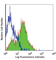
Monocyte-derived dendritic cells stained with HB15e PE
CD83 is a 43 kD single chain type I glycoprotein also known as HB15. A member of the immunoglobulin superfamily, CD83 is expressed on a subset of dendritic cells, Langerhans cells, and weakly on activated lymphocytes. Although CD83 is thought to play a role in antigen presentation and/or lymphocyte activation, the precise function of this protein is unknown. CD83 is considered to be a useful marker for mature dendritic cells.
Product DetailsProduct Details
- Reactivity
- Human
- Antibody Type
- Monoclonal
- Host Species
- Mouse
- Formulation
-
test sizes: Phosphate-buffered solution, pH 7.2, containing 0.09% sodium azide and BSA (origin USA).
µg size: Phosphate-buffered solution, pH 7.2, containing 0.09% sodium azide. - Preparation
- The antibody was purified by affinity chromatography, and conjugated with PE under optimal conditions.
- Concentration
- µg sizes: 0.2 mg/mLtest sizes: lot-specific (to obtain lot-specific concentration and expiration, please enter the lot number in our Certificate of Analysis online tool.)
- Storage & Handling
- The antibody solution should be stored undiluted between 2°C and 8°C, and protected from prolonged exposure to light. Do not freeze.
- Application
-
FC - Quality tested
- Recommended Usage
-
Each lot of this antibody is quality control tested by immunofluorescent staining with flow cytometric analysis. For flow cytometric staining, the suggested use of this reagent is 5 µl per million cells in 100 µl staining volume or 5 µl per 100 µl of whole blood.
- Excitation Laser
-
Blue Laser (488 nm)
Green Laser (532 nm)/Yellow-Green Laser (561 nm)
- Application Notes
-
Additional reported applications (for the relevant formats) include: immunohistochemical staining of acetone-fixed frozen tissue sections4.
-
Application References
(PubMed link indicates BioLegend citation) -
- Knapp W, et al. 1989. Leucocyte Typing IV. Oxford University Press New York.
- Zhou L, et al. 1995. J. Immunol. 154:3821.
- Cao W, et al. 2005. Biochem. J. 385:85.
- Lore K, et al. 2002. AIDS 16:683. (IHC)
- Cho H, et al. 2007. Physiol Genomics doi:10.1152/physiolgenomics.00051.2006
- Product Citations
- RRID
-
AB_314515 (BioLegend Cat. No. 305307)
AB_314516 (BioLegend Cat. No. 305308)
AB_2076529 (BioLegend Cat. No. 305322)
Antigen Details
- Structure
- Ig superfamily, single chain transmembrane glycoprotein, 43 kD
- Distribution
-
Dendritic cells, Langerhan cells, activated B and T cells
- Cell Type
- B cells, Dendritic cells, Langerhans cells, T cells
- Biology Area
- Costimulatory Molecules, Immunology
- Molecular Family
- CD Molecules
- Antigen References
-
1. Kozlow E, et al. 1993. Blood 81:454.
2. Zhou L, et al. 1992. J. Immunol. 149:735.
3. Zhou L, et al. 1995. Blood 86:3295. - Gene ID
- 9308 View all products for this Gene ID
- UniProt
- View information about CD83 on UniProt.org
Related Pages & Pathways
Pathways


Related FAQs
- What type of PE do you use in your conjugates?
- We use R-PE in our conjugates.
Customers Also Purchased
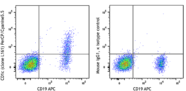
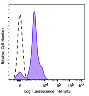
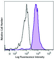
Compare Data Across All Formats
This data display is provided for general comparisons between formats.
Your actual data may vary due to variations in samples, target cells, instruments and their settings, staining conditions, and other factors.
If you need assistance with selecting the best format contact our expert technical support team.


Follow Us