 Login / Register


- Clone
- M5/114.15.2 (See other available formats)
- Regulatory Status
- RUO
- Other Names
- MHC class II
- Isotype
- Rat IgG2b, κ
- Ave. Rating
- Submit a Review
- Product Citations
- publications

-

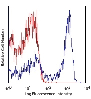
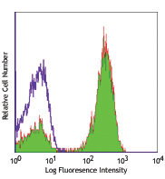
C57BL/6 mouse splenocytes were stained with anti-mouse I-A/I-E (clone M5/114.15.2)Alexa Fluor® 700 (filled histogram) or rat IgG2b, κ Alexa Fluor® 700 isotype control (open histogram). -

Confocal image of C57BL/6 mouse spleen sample acquired using the IBEX method of highly multiplexed antibody-based imaging: MHCII (blue) in Cycle 2 and F4/80 (magenta) in Cycle 2. Tissues were prepared using ~1% (vol/vol) formaldehyde and a detergent. Following fixation, samples are immersed in 30% (wt/vol) sucrose for cryoprotection. Images are courtesy of Drs. Andrea J. Radtke and Ronald N. Germain of the Center for Advanced Tissue Imaging (CAT-I) in the National Institute of Allergy and Infectious Diseases (NIAID, NIH). -

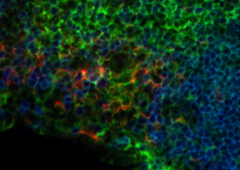
Confocal image of C57BL/6 mouse thymus sample acquired using the IBEX method of highly multiplexed antibody-based imaging: CD68 (cyan) in Cycle 3 and MHCII (red) in Cycle 3. Tissues were prepared using ~1% (vol/vol) formaldehyde and a detergent. Following fixation, samples are immersed in 30% (wt/vol) sucrose for cryoprotection. Images are courtesy of Drs. Andrea J. Radtke and Ronald N. Germain of the Center for Advanced Tissue Imaging (CAT-I) in the National Institute of Allergy and Infectious Diseases (NIAID, NIH). -

Confocal image of C57BL/6 mouse lung sample acquired using the IBEX method of highly multiplexed antibody-based imaging: EpCAM (blue) in Cycle 2, MHCII (magenta) in Cycle 2, and CD169 (green) in Cycle 4. Tissues were prepared using ~1% (vol/vol) formaldehyde and a detergent. Following fixation, samples are immersed in 30% (wt/vol) sucrose for cryoprotection. Images are courtesy of Drs. Andrea J. Radtke and Ronald N. Germain of the Center for Advanced Tissue Imaging (CAT-I) in the National Institute of Allergy and Infectious Diseases (NIAID, NIH). -

Confocal image of C57BL/6 mouse small intestine sample acquired using the IBEX method of highly multiplexed antibody-based imaging: EpCAM (blue) in Cycle 1, CD31 (magenta) in Cycle 2, and MHCII (green) in Cycle 3. Tissues were prepared using ~1% (vol/vol) formaldehyde and a detergent. Following fixation, samples are immersed in 30% (wt/vol) sucrose for cryoprotection. Images are courtesy of Drs. Andrea J. Radtke and Ronald N. Germain of the Center for Advanced Tissue Imaging (CAT-I) in the National Institute of Allergy and Infectious Diseases (NIAID, NIH). -

Confocal image of C57BL/6 mouse liver sample acquired using the IBEX method of highly multiplexed antibody-based imaging: E-Cadherin (blue) in Cycle 1 and MHCII (magenta) in Cycle 4. Tissues were prepared using ~1% (vol/vol) formaldehyde and a detergent. Following fixation, samples are immersed in 30% (wt/vol) sucrose for cryoprotection. Images are courtesy of Drs. Andrea J. Radtke and Ronald N. Germain of the Center for Advanced Tissue Imaging (CAT-I) in the National Institute of Allergy and Infectious Diseases (NIAID, NIH).
| Cat # | Size | Price | Quantity Check Availability | Save | ||
|---|---|---|---|---|---|---|
| 107621 | 25 µg | 76€ | ||||
| 107622 | 100 µg | 172€ | ||||
These class II molecules are expressed on antigen presenting cells (including B cells) and a subset of T cells from H-2b,d,q,r bearing mice and are involved in antigen presentation to T cells expressing CD3/TCR and CD4 proteins.
Product DetailsProduct Details
- Reactivity
- Mouse
- Antibody Type
- Monoclonal
- Host Species
- Rat
- Immunogen
- Activated C57BL/6 mouse spleen cells
- Formulation
- Phosphate-buffered solution, pH 7.2, containing 0.09% sodium azide.
- Preparation
- The antibody was purified by affinity chromatography and conjugated with Alexa Fluor® 700 under optimal conditions.
- Concentration
- 0.5 mg/ml
- Storage & Handling
- The antibody solution should be stored undiluted between 2°C and 8°C, and protected from prolonged exposure to light. Do not freeze.
- Application
-
FC - Quality tested
SB - Reported in the literature, not verified in house
- Recommended Usage
-
Each lot of this antibody is quality control tested by immunofluorescent staining with flow cytometric analysis. The suggested use of this reagent is ≤0.25 µg per million cells in 100 µl volume. It is highly recommended that the reagent be titrated for optimal performance for each application.
* Alexa Fluor® 700 has a maximum emission of 719 nm when it is excited at 633 nm / 635 nm. Prior to using Alexa Fluor® 700 conjugate for flow cytometric analysis, please verify your flow cytometer's capability of exciting and detecting the fluorochrome.
Alexa Fluor® and Pacific Blue™ are trademarks of Life Technologies Corporation.
View full statement regarding label licenses - Excitation Laser
-
Red Laser (633 nm)
- Application Notes
-
The M5/114.15.2 antibody reacts with a polymorphic determinant shared by the I-Ab, I-Ad, I-Aq, I-Ed, and I-Ek MHC class II alloantigens from mice carrying H-2p,r,q,b,d,u haplotypes. Clone M5/114.15.2 however does not react wtih I-Af, I-Ak, or I-As MHC class II alloantigens.1
Additional reported applications (for the relevant formats) include: immunoprecipitation1, immunohistochemistry of frozen sections2,3,6, in vitro and in vivo blocking of antigen presentation or ligand binding4-7, and spatial biology (IBEX)17,18. The Ultra-LEAF™ purified antibody (Endotoxin < 0.01 EU/µg, Azide-Free, 0.2 µm filtered) is recommended for functional assays (Cat. Nos. 107655 & 107656). - Additional Product Notes
-
Iterative Bleaching Extended multi-pleXity (IBEX) is a fluorescent imaging technique capable of highly-multiplexed spatial analysis. The method relies on cyclical bleaching of panels of fluorescent antibodies in order to image and analyze many markers over multiple cycles of staining, imaging, and, bleaching. It is a community-developed open-access method developed by the Center for Advanced Tissue Imaging (CAT-I) in the National Institute of Allergy and Infectious Diseases (NIAID, NIH).
- Application References
-
- Bhattacharya A, et al. 1981. J. Immunol. 127:2488. (IP)
- Viville S, et al. 1993. Cell 72:635. (IHC)
- Nelson AJ, et al. 1993. J. Immunol. 151:2453. (IHC)
- Shi Y, et al. 1998. J. Exp. Med. 187:367. (Block)
- Yamashita I, et al. 1993. Int. Immunol. 5:1139.
- Guo M, et al. 1995. Zygote 3:65. (IHC)
- Kim A, et al. 2004. Exp. Mol. Med. 36:428. (Block)
- Luckashenak NA, et al. 2006. J. Immunol. 177:5177.
- Venanzi ES, et al. 2007. J. Immunol. 179:5693.
- Christensen SR, et al. 2006. Immunity 25:417. PubMed
- Matte-Martone C, et al. 2008. Blood 111:3884. PubMed
- De Pascalis R, et al. 2008. Infect. Immun. 76:4311. PubMed
- Kuns RD, et al. 2009. Blood 113:5999. PubMed
- Sabatino JJ, et al. 2011. J. Exp. Med. 208:81. PubMed
- Draber P, et al. 2011. Mol Cell Biol. 22:4550. PubMed
- Fu H, et al. 2014. Nat Commun. 5:3436. PubMed
- Radtke AJ, et al. 2020. Proc Natl Acad Sci U S A. 117:33455-65. (SB) PubMed
- Radtke AJ, et al. 2022. Nat Protoc. 17:378-401. (SB) PubMed
- Product Citations
- RRID
-
AB_493726 (BioLegend Cat. No. 107621)
AB_493727 (BioLegend Cat. No. 107622)
Antigen Details
- Structure
- MHC class II
- Distribution
-
B cell and activated T cells, APCs of the H-2b,d,q,r bearing mice
- Function
- Antigen presentation
- Ligand/Receptor
- CD3/TCR, CD4
- Cell Type
- Antigen-presenting cells, B cells, Dendritic cells, T cells, Tregs
- Biology Area
- Immunology, Innate Immunity
- Molecular Family
- MHC Antigens
- Antigen References
-
1. Watts C. 1997. Ann. Rev. Immunol. 15:821.
2. Pamer E, et al. 1998. Ann. Rev. Immunol. 16:323. - Gene ID
- 14961 View all products for this Gene ID 14969 View all products for this Gene ID
- UniProt
- View information about I-A/I-E on UniProt.org
Related Pages & Pathways
Pathways


Related FAQs
- If an antibody clone has been previously successfully used in IBEX in one fluorescent format, will other antibody formats work as well?
-
It’s likely that other fluorophore conjugates to the same antibody clone will also be compatible with IBEX using the same sample fixation procedure. Ultimately a directly conjugated antibody’s utility in fluorescent imaging and IBEX may be specific to the sample and microscope being used in the experiment. Some antibody clone conjugates may perform better than others due to performance differences in non-specific binding, fluorophore brightness, and other biochemical properties unique to that conjugate.
- Will antibodies my lab is already using for fluorescent or chromogenic IHC work in IBEX?
-
Fundamentally, IBEX as a technique that works much in the same way as single antibody panels or single marker IF/IHC. If you’re already successfully using an antibody clone on a sample of interest, it is likely that clone will have utility in IBEX. It is expected some optimization and testing of different antibody fluorophore conjugates will be required to find a suitable format; however, legacy microscopy techniques like chromogenic IHC on fixed or frozen tissue is an excellent place to start looking for useful antibodies.
- Are other fluorophores compatible with IBEX?
-
Over 18 fluorescent formats have been screened for use in IBEX, however, it is likely that other fluorophores are able to be rapidly bleached in IBEX. If a fluorophore format is already suitable for your imaging platform it can be tested for compatibility in IBEX.
- The same antibody works in one tissue type but not another. What is happening?
-
Differences in tissue properties may impact both the ability of an antibody to bind its target specifically and impact the ability of a specific fluorophore conjugate to overcome the background fluorescent signal in a given tissue. Secondary stains, as well as testing multiple fluorescent conjugates of the same clone, may help to troubleshoot challenging targets or tissues. Using a reference control tissue may also give confidence in the specificity of your staining.
- How can I be sure the staining I’m seeing in my tissue is real?
-
In general, best practices for validating an antibody in traditional chromogenic or fluorescent IHC are applicable to IBEX. Please reference the Nature Methods review on antibody based multiplexed imaging for resources on validating antibodies for IBEX.
Customers Also Purchased
Compare Data Across All Formats
This data display is provided for general comparisons between formats.
Your actual data may vary due to variations in samples, target cells, instruments and their settings, staining conditions, and other factors.
If you need assistance with selecting the best format contact our expert technical support team.


Follow Us