 Login / Register


- Clone
- GoH3 (See other available formats)
- Regulatory Status
- RUO
- Workshop
- HCDM listed
- Other Names
- VLA-6 α chain, α6 integrin, integrin α6, ITGA6
- Isotype
- Rat IgG2a, κ
- Ave. Rating
- Submit a Review
- Product Citations
- publications

-

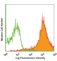
Human peripheral blood lymphocytes stained with GoH3 PE
| Cat # | Size | Price | Quantity Check Availability | Save | ||
|---|---|---|---|---|---|---|
| 313611 | 25 tests | 120€ | ||||
| 313612 | 100 tests | 173€ | ||||
CD49f is a 120 kD integrin family member also known as VLA-6 α chain and α6 integrin subunit. CD49f associates with either integrin β1 (CD29) or integrin β4 (CD104) to form receptors (VLA-6 or α6β4 complex) for laminin and kalinin. CD49f is expressed on platelets, monocytes, T cells, placental trophoblasts, and epithelial and endothelial cells. CD49f is involved in adhesion and can act as a co-stimulatory molecule for T cell activation and proliferation.
Product DetailsProduct Details
- Reactivity
- Human,Mouse,Cynomolgus,Rhesus
- Antibody Type
- Monoclonal
- Host Species
- Rat
- Immunogen
- Mouse mammary tumor cells
- Formulation
- Phosphate-buffered solution, pH 7.2, containing 0.09% sodium azide and BSA (origin USA)
- Preparation
- The antibody was purified by affinity chromatography, and conjugated with PE under optimal conditions.
- Concentration
- Lot-specific (to obtain lot-specific concentration and expiration, please enter the lot number in our Certificate of Analysis online tool.)
- Storage & Handling
- The antibody solution should be stored undiluted between 2°C and 8°C, and protected from prolonged exposure to light. Do not freeze.
- Application
-
FC - Quality tested
- Recommended Usage
-
Each lot of this antibody is quality control tested by immunofluorescent staining with flow cytometric analysis. For flow cytometric staining, the suggested use of this reagent is 5 µl per million cells in 100 µl staining volume or 5 µl per 100 µl of whole blood.
- Excitation Laser
-
Blue Laser (488 nm)
Green Laser (532 nm)/Yellow-Green Laser (561 nm)
- Application Notes
-
Additional reported applications (for the relevant formats) include: immunoprecipitation1,5, in vitro and in vivo blocking of cell binding to laminin and blocking the function of integrin a61,4, and immunohistochemistry of acetone-fixed frozen sections2,3,5. The GoH3 antibody has been reported to block laminin binding in vitro and to block integrin a6 function in vivo.
- Application References
-
- Georas SN, et al. 1993. Blood 82:2872. (IP, Block)
- Honda T, et al. 1995. J. Clin. Endocrinol. Metab. 80:2899. (IHC)
- Sonnenberg A, et al. 1986. J. Histochem. Cytochem. 34:1037. (IHC)
- Nakamura K, et al. 1997 Biochem. Biophys. Res. Commun. 235:524. (Block)
- Sonnenberg A, et al. 1987 J. Biol. Chem. 262:10376. (IP, IHC)
- Deregibus MC, et al. 2007. Blood doi:10.1182/blood-2007-03-078709.
- Horwitz KB, et al. 2008. Proc Natl Acad Sci USA. 105:5774. PubMed
- Nardella C, et al. 2009. Sci Signal. 2:55. PubMed
- Xu T, et al. 2010. Mol Cancer Ther. 9:438. PubMed
- Stepp MA, et al. 2007. J Cell Sci. 120:2851. PubMed
- Jo M, et al. 2010. Cancer Res. 70:8948. PubMed
- Yoshino N, et al. 2000. Exp. Anim. (Tokyo) 49:97. (FC)
- Grange C, et al. 2011. Cancer Res. 71:5346. PubMed
- Lai KP, et al. 2012. Mol Endocrinol. 26:52. PubMed
- Oeztuerk-Winder F, et al. 2012. EMBO J. 31:3431. (FC) PubMed
- Product Citations
- RRID
-
AB_893374 (BioLegend Cat. No. 313611)
AB_893373 (BioLegend Cat. No. 313612)
Antigen Details
- Structure
- Integrin family, associates with β1 or β4, 120 kD
- Distribution
-
Platelets, monocytes, T cells, placental trophoblasts, epithelial and endothelial cells
- Function
- Adhesion, receptor for laminin and kalinin; laminin binding to VLA-6 induces T cell co-stimulation for proliferation and activation
- Ligand/Receptor
- With integrin β1 (CD29) forms VLA-6, with integrin β4 (CD104) forms a6β4 integrin; laminin and kalinin are ligands for these receptors
- Cell Type
- Embryonic Stem Cells, Endothelial cells, Epithelial cells, Monocytes, Platelets, T cells
- Biology Area
- Cell Adhesion, Cell Biology, Immunology, Innate Immunity, Stem Cells
- Molecular Family
- Adhesion Molecules, CD Molecules
- Antigen References
-
1. Sonnenberg A, et al. 1990. J. Cell Biol. 110:2145.
2. Sonnenberg A, et al. 1990. J. Cell. Sci. 96:207.
3. Aumailley M, et al. 1990. Exp. Cell Res. 188:55.
4. Niessen CM, et al. 1994. Exp. Cell Res. 211:360. - Gene ID
- 16403 View all products for this Gene ID 3655 View all products for this Gene ID
- UniProt
- View information about CD49f on UniProt.org
Related Pages & Pathways
Pathways


Related FAQs
- What type of PE do you use in your conjugates?
- We use R-PE in our conjugates.
Customers Also Purchased
Compare Data Across All Formats
This data display is provided for general comparisons between formats.
Your actual data may vary due to variations in samples, target cells, instruments and their settings, staining conditions, and other factors.
If you need assistance with selecting the best format contact our expert technical support team.


Follow Us