 Login / Register


- Clone
- 3/23 (See other available formats)
- Regulatory Status
- RUO
- Other Names
- Bp50, TNFRSF5
- Isotype
- Rat IgG2a, κ
- Ave. Rating
- Submit a Review
- Product Citations
- publications

-

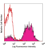
BALB/c splenocytes stained with 3/23 PE
| Cat # | Size | Price | Quantity Check Availability | Save | ||
|---|---|---|---|---|---|---|
| 124609 | 25 µg | 72€ | ||||
| 124610 | 100 µg | 220€ | ||||
CD40 is a 48 kD type I transmembrane glycoprotein also known as Bp50. It is a member of the tumor necrosis factor receptor (TNFR) superfamily and is expressed on B cells, basal epithelial cells, macrophages, follicular dendritic cells, endothelial cells, and a subset of CD34+ hematopoietic progenitors. CD40 regulates B cell development/maturation, Ig isotype switching and, in combination with other signals such as IL-4, protects B cells from surface Ig-induced apoptosis and promotes proliferation. Interaction of CD40 with its ligand CD154 (gp39), which is expressed on activated T cells, is important in costimulation and immune regulation.
Product DetailsProduct Details
- Reactivity
- Mouse
- Antibody Type
- Monoclonal
- Host Species
- Rat
- Immunogen
- Recombinant mouse CD40 protein
- Formulation
- Phosphate-buffered solution, pH 7.2, containing 0.09% sodium azide.
- Preparation
- The antibody was purified by affinity chromatography, and conjugated with PE under optimal conditions.
- Concentration
- 0.2 mg/ml
- Storage & Handling
- The antibody solution should be stored undiluted between 2°C and 8°C, and protected from prolonged exposure to light. Do not freeze.
- Application
-
FC - Quality tested
- Recommended Usage
-
Each lot of this antibody is quality control tested by immunofluorescent staining with flow cytometric analysis. For flow cytometric staining, the suggested use of this reagent is ≤1.0 µg per million cells in 100 µl volume. It is recommended that the reagent be titrated for optimal performance for each application.
- Excitation Laser
-
Blue Laser (488 nm)
Green Laser (532 nm)/Yellow-Green Laser (561 nm)
- Application Notes
-
For highly sensitive assays, we recommend Ultra-LEAF™ purified antibody (Cat. No. 124627 &124628) with a low endotoxin limit (Endotoxin < 0.01 EU/µg).
- Application References
-
- Hasbold J, et al. 1994. Eur. J. Immunol. 24:1835.
- Bourgeois C, et al. 2002. Science 297:2060.
- Product Citations
- RRID
-
AB_1134084 (BioLegend Cat. No. 124609)
AB_1134075 (BioLegend Cat. No. 124610)
Antigen Details
- Structure
- 48 kD type I transmembrane glycoprotein, a member of the tumor necrosis factor receptor (TNFR) superfamily.
- Distribution
-
Expressed on B lymphocytes and other antigen-presenting cells, such as macrophages, follicular dendritic cells, etc.
- Function
- Regulates B cell growth and differentiation and Ig isotype switching. Interaction of CD40 with its ligand, CD154, on activated T cells is important for immune response.
- Ligand/Receptor
- CD154 on T cells
- Cell Type
- Antigen-presenting cells, B cells, Dendritic cells, Macrophages
- Biology Area
- Cell Biology, Costimulatory Molecules, Immunology, Neuroscience, Neuroscience Cell Markers
- Molecular Family
- CD Molecules
- Antigen References
-
1. Grewal IS, et al. 1998. Annu Rev Immunol 16:111.
- Gene ID
- 21939 View all products for this Gene ID
- UniProt
- View information about CD40 on UniProt.org
Related Pages & Pathways
Pathways


Related FAQs
- What type of PE do you use in your conjugates?
- We use R-PE in our conjugates.
Customers Also Purchased
Compare Data Across All Formats
This data display is provided for general comparisons between formats.
Your actual data may vary due to variations in samples, target cells, instruments and their settings, staining conditions, and other factors.
If you need assistance with selecting the best format contact our expert technical support team.


Follow Us