 Login / Register


- Clone
- D7 (See other available formats)
- Regulatory Status
- RUO
- Other Names
- Sca-1
- Isotype
- Rat IgG2a, κ
- Ave. Rating
- Submit a Review
- Product Citations
- publications

-

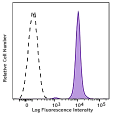
C57BL/6 mouse splenocytes stained with D7 PE/Cyanine7
| Cat # | Size | Price | Quantity Check Availability | Save | ||
|---|---|---|---|---|---|---|
| 108113 | 25 µg | 82€ | ||||
| 108114 | 100 µg | 253€ | ||||
Ly-6A/E, also known as Sca-1, is an 18 kD member of the Ly-6 multigene family. Ly6A/E is a glycosylphosphatidylinositol (GPI)-linked protein expressed on hematopoietic stem cells. In mice expressing the Ly-6.2 haplotype (e.g., AKR, C57BL, C57BR, DBA/2, SJL, SWR, and 129), Ly-6A/E is also expressed on peripheral B lymphocytes and thymic and peripheral T lymphocytes. Strains expressing the Ly-6.1 haplotype (e.g., BALB/c, CBA, C3H/He, DBA/1, and NZB) have low Ly-6A/E expression on resting peripheral lymphocytes. The expression of Ly-6A/E on lymphocytes is upregulated upon activation from both Ly6.1 and Ly6.2 haplotype mice. Ly-6A/E is thought to be involved in the regulation of both T and B cell responses.
Product DetailsProduct Details
- Reactivity
- Mouse
- Antibody Type
- Monoclonal
- Host Species
- Rat
- Immunogen
- IL-2-dependent mouse T-cell line (CTL-L)
- Formulation
- Phosphate-buffered solution, pH 7.2, containing 0.09% sodium azide.
- Preparation
- The antibody was purified by affinity chromatography, and conjugated with PE/Cyanine7 under optimal conditions.
- Concentration
- 0.2 mg/ml
- Storage & Handling
- The Ly-6A/E antibody solution should be stored undiluted between 2°C and 8°C, and protected from prolonged exposure to light. Do not freeze.
- Application
-
FC - Quality tested
- Recommended Usage
-
Each lot of this antibody is quality control tested by immunofluorescent staining with flow cytometric analysis. For flow cytometric staining, the suggested use of this reagent is ≤ 0.25 µg per 106 cells in 100 µl volume. It is recommended that the reagent be titrated for optimal performance for each application.
- Excitation Laser
-
Blue Laser (488 nm)
Green Laser (532 nm)/Yellow-Green Laser (561 nm)
- Application Notes
-
The D7 antibody has been reported to induce T cell activation and inhibit TCR-induced IL-2 production. Additional reported applications (for the relevant formats) include: Western blotting1,2, immunoprecipitation1, in vitro lymphocyte activation3-6, induction of redirected lysis7, induction of T cell inhibitory signalling8, immunofluorescence9, and immunohistochemical staining of acetone-fixed frozen sections13 and Bouin-fixed, paraffin-embedded samples9.
The two Sca-1 recognizing clones D7 and E13-161.7 have been shown to bind distinct epitopes due to the inability of D7 to block the binding of E13-161.7.14 - Additional Product Notes
- BioLegend is in the process of converting the name PE/Cy7 to PE/Cyanine7. The dye molecule remains the same, so you should expect the same quality and performance from our PE/Cyanine7 products. Please contact Technical Service if you have any questions.
- Application References
-
- Ortega G, et al. 1986. J. Immunol. 137:3240. (WB, IP)
- Palfree RGE, et al. 1986. Immunogenetics 23:197. (WB)
- Codias EK, et al. 1990. J. Immunol. 144:2197.
- Malek TR, et al. 1986. J. Exp. Med. 164:709.
- Codias EK, et al. 1990. J. Immunol. 145:1407.
- Ivanov V, et al. 1994. J. Immunol. 153:2394.
- Karlhofer FM, et al. 1991. J. Immunol. 146:3662.
- Fleming T, et al. 1994. J. Immunol. 153:1955.
- van Bragt MPA, et al. 2005. Biol. Reprod. 73:634. (IF, IHC)
- Umland O, et al. 2007. J. Immunol. 178:4147.
- Cridland SO, et al. 2009. Blood Cell. Mol. Dis. 45:149. (FC) PubMed
- Pronk CJ, et al. 2011. J. Exp Med. PubMed
- English A, et al. 2000. J. Immunol. 165:3763. (IHC)
- Bamezai A and Rock KL. 1995. Proc. Natl. Acad. Sci. USA 92:4294.
- Wiesner DL, et al. 2015. PLoS Pathog. 11:1004701. PubMed
- Product Citations
- RRID
-
AB_493597 (BioLegend Cat. No. 108113)
AB_493596 (BioLegend Cat. No. 108114)
Antigen Details
- Structure
- Ly-6 multigene family, 18 kD
- Distribution
-
Hematopoietic stem cells, activated T cells and B cells, subset of resting B cells and T cells
- Function
- Regulates B and T cell responses
- Cell Type
- B cells, Hematopoietic stem and progenitors, Mesenchymal Stem Cells, T cells
- Biology Area
- Immunology, Stem Cells
- Antigen References
-
1. Rock KL, et al. 1989. Immunol. Rev. 111:195.
2. Morrison SJ, et al. 1994. Immunity 1:661.
3. Spangrude GJ, et al. 1988. J. Immunol. 141:3697.
4. Malek T, et al. 1986. J. Exp. Med. 164:709. - Gene ID
- 110454 View all products for this Gene ID
- UniProt
- View information about Ly-6A/E on UniProt.org


Customers Also Purchased
Compare Data Across All Formats
This data display is provided for general comparisons between formats.
Your actual data may vary due to variations in samples, target cells, instruments and their settings, staining conditions, and other factors.
If you need assistance with selecting the best format contact our expert technical support team.


Follow Us