 Login / Register


- Clone
- 10.1 (See other available formats)
- Regulatory Status
- RUO
- Workshop
- VI MA36
- Other Names
- FcγRI, FcR I
- Isotype
- Mouse IgG1, κ
- Ave. Rating
- Submit a Review
- Product Citations
- publications

-

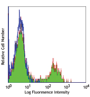
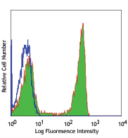
Human peripheral blood monocytes were stained with CD64 (clone 10.1) Purified (filled histogram) or Purified Mouse IgG1, κ isotype control (open histogram) followed by anti-mouse IgG FITC
| Cat # | Size | Price | Quantity Check Availability | Save | ||
|---|---|---|---|---|---|---|
| 305002 | 100 µg | 82€ | ||||
CD64 is a 72 kD single chain type I glycoprotein also known as FcγRI and FcR I. CD64 is a member of the immunoglobulin superfamily and is expressed on monocytes/macrophages, dendritic cells, and activated granulocytes. The expression can be upregulated by IFN-γ stimulation. CD64 binds IgG immune complex. It plays a role in antigen capture, phagocytosis of IgG/antigen complexes, and antibody-dependent cellular cytotoxicity (ADCC).
Product DetailsProduct Details
- Reactivity
- Human,Cynomolgus,Rhesus
- Antibody Type
- Monoclonal
- Host Species
- Mouse
- Immunogen
- Human rheumatoid synovial fluid cells and fibronectin-purified monocytes.
- Formulation
- Phosphate-buffered solution, pH 7.2, containing 0.09% sodium azide.
- Preparation
- The antibody was purified by affinity chromatography.
- Concentration
- 0.5 mg/ml
- Storage & Handling
- The antibody solution should be stored undiluted between 2°C and 8°C.
- Application
-
FC - Quality tested
IHC-F, Block - Reported in the literature, not verified in house - Recommended Usage
-
Each lot of this antibody is quality control tested by immunofluorescent staining with flow cytometric analysis. For flow cytometric staining, the suggested use of this reagent is ≤ 1.0 µg per 106 cells in 100 µl volume or 100 µl of whole blood. It is recommended that the reagent be titrated for optimal performance for each application.
- Application Notes
-
Clone 10.1 recognizes the EC3 epitope of CD64. While both contain the EC3 domain, in-house testing suggests that clone 10.1 preferentially binds to CD64A (FcγRIA), but not CD64B (FcγRIB). Additional reported applications (for the relevant formats) include: blocking of human IgG3 and murine IgG2a binding to FcγRI2,5,6,11 and immunohistochemical staining of acetone-fixed frozen tissue sections12.
- Application References
-
- McMichael A, et al. Eds. 1987. Leucocyte Typing III. Oxford University Press. New York.
- Schlossman S, et al. Eds. 1995. Leucocyte Typing V. Oxford University Press. New York. p. 874.
- Kishimoto T, et al. Eds. 1997. Leucocyte Typing VI. Garland Publishing Inc. London.
- Holl V, et al. 2004. J. Immunol. 173:6274.
- Hober D, et al. 2002. J. Gen. Virol. 83:2169.
- Cho HJ, et al. 2007. Physiol Genomics 149:60.
- van Tits L, et al. 2005. Arterioscler Thromb Vasc Biol. 25:717. PubMed
- Bruhns P, et al. 2008. Blood 113:3716. PubMed
- Yoshino N, et al. 2000. Exp. Anim. (Tokyo) 49:97. (FC)
- Carter DL, et al. 1999. Cytometry 37:41. (FC)
- Dougherty GJ, et al. 1987. Eur. J. Immunol. 17:1453.
- Blom AB, et al. 2003. Arthritis Rheum. 48(4):1002-14. (IHC)
- Product Citations
- RRID
-
AB_314486 (BioLegend Cat. No. 305002)
Antigen Details
- Structure
- Ig superfamily, type I glycoprotein, 72 kD
- Distribution
-
Monocytes, macrophages, dendritic cells, activated granulocytes
- Function
- Phagocytosis, ADCC
- Ligand/Receptor
- IgG receptor
- Cell Type
- Dendritic cells, Granulocytes, Macrophages, Monocytes
- Biology Area
- Immunology, Innate Immunity
- Molecular Family
- CD Molecules, Fc Receptors
- Antigen References
-
1. Hulett M, et al. 1994. Adv. Immunol. 57:1.
2. van de Winkel J, et al. 1993. Immunol. Today 14:215. - Gene ID
- 2209 View all products for this Gene ID
- UniProt
- View information about CD64 on UniProt.org


Related FAQs
- Is our human Trustain FcX™ (cat# 422302) compatible with anti human CD16, CD32 and CD64 clones 3G8, FUN-2 and 10.1 respectively?
-
Yes
Customers Also Purchased
Compare Data Across All Formats
This data display is provided for general comparisons between formats.
Your actual data may vary due to variations in samples, target cells, instruments and their settings, staining conditions, and other factors.
If you need assistance with selecting the best format contact our expert technical support team.


Follow Us