 Login / Register


- Clone
- 53-6.7 (See other available formats)
- Regulatory Status
- RUO
- Other Names
- T8, Lyt2, Ly-2
- Isotype
- Rat IgG2a, κ
- Ave. Rating
- Submit a Review
- Product Citations
- publications

-

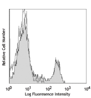
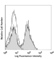
C57BL/6 mouse splenocytes were stained with LEAF™ purified CD8 (clone 53-6.7) (filled histogram) or rat IgG2a, κ isotype control (open histogram), followed by anti-rat IgG FITC.
Select size of product is eligible for a 40% discount! Promotion valid until September 30, 2024. Exclusions apply. To view full promotion terms and conditions or to contact your local BioLegend representative to receive a quote, visit our webpage.
CD8, also known as Lyt-2, Ly-2, or T8, consists of disulfide-linked α and β chains that form the α(CD8a)/β(CD8b) heterodimer and α/α homodimer. CD8a is a 34 kD protein that belongs to the immunoglobulin family. The CD8 α/β heterodimer is expressed on the surface of most thymocytes and a subset of mature TCR α/β T cells. CD8 expression on mature T cells is non-overlapping with CD4. The CD8 α/α homodimer is expressed on a subset of γ/δ TCR-bearing T cells, NK cells, intestinal intraepithelial lymphocytes, and lymphoid dendritic cells. CD8 is an antigen co-receptor on T cells that interacts with MHC class I on antigen-presenting cells or epithelial cells. CD8 promotes T cell activation through its association with the TCR complex and protein tyrosine kinase lck.
Product DetailsProduct Details
- Reactivity
- Mouse
- Antibody Type
- Monoclonal
- Host Species
- Rat
- Immunogen
- Mouse thymus or spleen
- Formulation
- 0.2 µm filtered in phosphate-buffered solution, pH 7.2, containing no preservative.
- Preparation
- The Ultra-LEAF™ (Low Endotoxin, Azide-Free) antibody was purified by affinity chromatography.
- Concentration
- The antibody is bottled at the concentration indicated on the vial, typically between 2 mg/mL and 3 mg/mL. Older lots may have also been bottled at 1 mg/mL. To obtain lot-specific concentration and expiration, please enter the lot number in our Certificate of Analysis online tool.
- Storage & Handling
- The antibody solution should be stored undiluted between 2°C and 8°C. This Ultra-LEAF™ solution contains no preservative; handle under aseptic conditions.
- Application
-
FC - Quality tested
CyTOF® - Verified
IHC, IP, Depletion, Block - Reported in the literature, not verified in house - Recommended Usage
-
Each lot of this antibody is quality control tested by immunofluorescent staining with flow cytometric analysis. For flow cytometric staining, the suggested use of this reagent is ≤0.25 µg per million cells in 100 µl volume or 100 µl of whole blood. It is recommended that the reagent be titrated for optimal performance for each application.
- Application Notes
-
Clone 53-6.7 antibody competes with clone 5H10-1 antibody for binding to thymocytes3. The 53-6.7 antibody has been reported to block antigen presentation via MHC class I and inhibit T cell responses to IL-2. This antibody has also been used for depletion of CD8a+ cells. Additional reported applications (for the relevant formats) include: immunoprecipitation1,3, in vivo and in vitro cell depletion2,10,15, inhibition of CD8 T cell proliferation3, blocking of cytotoxicity3,4, immunohistochemical staining5,6 of acetone-fixed frozen sections and zinc-fixed paraffin-embedded sections, and spatial biology (IBEX)29,30. Clone 53-6.7 is not recommended for immunohistochemistry of formalin-fixed paraffin sections. The Ultra-LEAF™ purified antibody (Endotoxin < 0.01 EU/µg, Azide-Free, 0.2 µm filtered) is recommended for functional assays or in vivo studies (Cat No. 100746).
- Application References
-
- Ledbetter JA, et al. 1979. Immunol. Rev. 47:63. (IHC, IP)
- Hathcock KS. 1991. Current Protocols in Immunology. 3.4.1. (Deplete)
- Takahashi K, et al. 1992. P. Natl. Acad. Sci. USA 89:5557. (Block, IP)
- Ledbetter JA, et al. 1981. J. Exp. Med. 153:1503. (Block)
- Hata H, et al. 2004. J. Clin. Invest. 114:582. (IHC)
- Fan WY, et al. 2001. Exp. Biol. Med. 226:1045. (IHC)
- Shih FF, et al. 2006. J. Immunol. 176:3438. (FC)
- Kamimura D, et al. 2006. J. Immunol. 177:306.
- Bouwer HGA, et al. 2006. P. Natl. Acad. Sci. USA 103:5102. (FC, Deplete)
- Kao C, et al. 2005. Int. Immunol. 17:1607. PubMed
- Ko SY, et al. 2005. J. Immunol. 175:3309. (FC) PubMed
- Rasmussen JW, et al. 2006. Infect. Immun. 74:6590. PubMed
- Lee CH, et al. 2009. Clin. Cancer Res. PubMed
- Geiben-Lynn R, et al. 2008. Blood 112:4585. (Deplete) PubMed
- Kingeter LM, et al. 2008. J. Immunol. 181:6244. PubMed
- Guo Y, et al. 2008. Blood 112:480. PubMed
- Andrews DM, et al. 2008. J. Virol. 82:4931. PubMed
- Britschqui MR, et al. 2008. J. Immunol. 181:7681. PubMed
- Kenna TJ, et al. 2008. Blood 111:2091. PubMed
- Jordan JM, et al. 2008. Infect. Immun. 76:3717. PubMed
- Todd DJ, et al. 2009. J. Exp. Med. 206:2151. PubMed
- Bankoti J, et al. 2010. Toxicol. Sci. 115:422. (FC) PubMed
- Medyouf H, et al. 2010. Blood 115:1175. PubMed
- Riedl P, et al. 2009. J. Immunol. 183:370. PubMed
- Apte SH, et al. 2010. J. Immunol. 185:998. PubMed
- Bankoti J, et al. 2010. Toxicol. Sci. 115:422. (FC) PubMed
- del Rio ML, et al. 2011. Transpl. Int. 24:501. (FC) PubMed
- Cui L, et al. 2015. J Control Release. 206:220. PubMed
- Radtke AJ, et al. 2020. Proc Natl Acad Sci U S A. 117:33455-65. (SB) PubMed
- Radtke AJ, et al. 2022. Nat Protoc. 17:378-401. (SB) PubMed
- Product Citations
- RRID
-
AB_2810325 (BioLegend Cat. No. 100775)
AB_11147171 (BioLegend Cat. No. 100746)
AB_2810323 (BioLegend Cat. No. 100763)
AB_2810324 (BioLegend Cat. No. 100764)
AB_2810326 (BioLegend Cat. No. 100776)
AB_2810327 (BioLegend Cat. No. 100777)
Antigen Details
- Structure
- Ig superfamily, CD8α chain, 34 kD
- Distribution
-
Most thymocytes, T cell subset, some NK cells, lymphoid dendritic cells
- Function
- Co-receptor for TCR
- Ligand/Receptor
- MHC class I molecule
- Antigen References
-
1. Barclay A, et al. 1997. The Leukocyte Antigen FactsBook Academic Press.
2. Zamoyska R. 1994. Immunity 1:243.
3. Ellmeier W, et al. 1999. Annu. Rev. Immunol. 17:523. - Gene ID
- 12525 View all products for this Gene ID
- UniProt
- View information about CD8alpha on UniProt.org


Related FAQs
- Do you guarantee that your antibodies are totally pathogen free?
-
BioLegend does not test for pathogens in-house aside from the GoInVivo™ product line. However, upon request, this can be tested on a custom basis with an outside, independent laboratory.
- Does BioLegend test each Ultra-LEAF™ antibody by functional assay?
-
No, BioLegend does not test Ultra-LEAF™ antibodies by functional assays unless otherwise indicated. Due to the possible complexities and variations of uses of biofunctional antibodies in different assays and because of the large product portfolio, BioLegend does not currently perform functional assays as a routine QC for the antibodies. However, we do provide references in which the antibodies were used for functional assays and we do perform QC to verify the specificity and quality of the antibody based on our strict specification criteria.
- Does BioLegend test each Ultra-LEAF™ antibody for potential pathogens?
-
No, BioLegend does not test for pathogens in-house unless otherwise indicated. However, we can recommend an outside vendor to perform this testing as needed.
- Have you tested this Ultra-LEAF™ antibody for in vivo or in vitro applications?
-
We don't test our antibodies for in vivo or in vitro applications unless otherwise indicated. Depending on the product, the TDS may describe literature supporting usage of a particular product for bioassay. It may be best to further consult the literature to find clone specific information.
Customers Also Purchased
Compare Data Across All Formats
This data display is provided for general comparisons between formats.
Your actual data may vary due to variations in samples, target cells, instruments and their settings, staining conditions, and other factors.
If you need assistance with selecting the best format contact our expert technical support team.


Follow Us