 Login / Register


- Clone
- M5/114.15.2 (See other available formats)
- Regulatory Status
- RUO
- Other Names
- MHC class II
- Isotype
- Rat IgG2b, κ
- Ave. Rating
- Submit a Review
- Product Citations
- publications

-

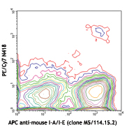
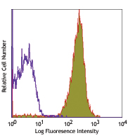
C57BL/6 mouse splenocytes were stained with anti-mouse CD11b FITC and I-A/I-E (clone M5/114.15.2) APC/Cyanine7 (left) or Rat IgG2b, κ APC/Cyanine7 isotype control (right).
| Cat # | Size | Price | Quantity Check Availability | Save | ||
|---|---|---|---|---|---|---|
| 107627 | 25 µg | 76€ | ||||
| 107628 | 100 µg | 234€ | ||||
These class II molecules are expressed on antigen presenting cells (including B cells) and a subset of T cells from H-2b,d,q,r bearing mice and are involved in antigen presentation to T cells expressing CD3/TCR and CD4 proteins.
Product DetailsProduct Details
- Verified Reactivity
- Mouse
- Antibody Type
- Monoclonal
- Host Species
- Rat
- Immunogen
- Activated C57BL/6 mouse spleen cells
- Formulation
- Phosphate-buffered solution, pH 7.2, containing 0.09% sodium azide.
- Preparation
- The antibody was purified by affinity chromatography, and conjugated with APC/Cyanine7 under optimal conditions.
- Concentration
- 0.2 mg/ml
- Storage & Handling
- The antibody solution should be stored undiluted between 2°C and 8°C, and protected from prolonged exposure to light. Do not freeze.
- Application
-
FC - Quality tested
- Recommended Usage
-
Each lot of this antibody is quality control tested by immunofluorescent staining with flow cytometric analysis. For flow cytometric staining, the suggested use of this reagent is = 0.25 µg per 106 cells in 100 µl volume. It is recommended that the reagent be titrated for optimal performance for each application.
- Excitation Laser
-
Red Laser (633 nm)
- Application Notes
-
The M5/114.15.2 antibody reacts with a polymorphic determinant shared by the I-Ab, I-Ad, I-Aq, I-Ed, and I-Ek MHC class II alloantigens from mice carrying H-2p,r,q,b,d,u haplotypes. Clone M5/114.15.2 however does not react wtih I-Af, I-Ak, or I-As MHC class II alloantigens.1
Additional reported applications (for the relevant formats) include: immunoprecipitation1, immunohistochemistry of frozen sections2,3,6, in vitro and in vivo blocking of antigen presentation or ligand binding4-7, and spatial biology (IBEX)17,18. The Ultra-LEAF™ purified antibody (Endotoxin < 0.01 EU/µg, Azide-Free, 0.2 µm filtered) is recommended for functional assays (Cat. Nos. 107655 & 107656). - Additional Product Notes
- BioLegend is in the process of converting the name APC/Cy7 to APC/Cyanine7. The dye molecule remains the same, so you should expect the same quality and performance from our APC/Cyanine7 products. Please contact Technical Service if you have any questions.
- Application References
-
- Bhattacharya A, et al. 1981. J. Immunol. 127:2488. (IP)
- Viville S, et al. 1993. Cell 72:635. (IHC)
- Nelson AJ, et al. 1993. J. Immunol. 151:2453. (IHC)
- Shi Y, et al. 1998. J. Exp. Med. 187:367. (Block)
- Yamashita I, et al. 1993. Int. Immunol. 5:1139.
- Guo M, et al. 1995. Zygote 3:65. (IHC)
- Kim A, et al. 2004. Exp. Mol. Med. 36:428. (Block)
- Luckashenak NA, et al. 2006. J. Immunol. 177:5177.
- Venanzi ES, et al. 2007. J. Immunol. 179:5693.
- Christensen SR, et al. 2006. Immunity 25:417. PubMed
- Matte-Martone C, et al. 2008. Blood 111:3884. PubMed
- De Pascalis R, et al. 2008. Infect. Immun. 76:4311. PubMed
- Kuns RD, et al. 2009. Blood 113:5999. PubMed
- Sabatino JJ, et al. 2011. J. Exp. Med. 208:81. PubMed
- Draber P, et al. 2011. Mol Cell Biol. 22:4550. PubMed
- Fu H, et al. 2014. Nat Commun. 5:3436. PubMed
- Radtke AJ, et al. 2020. Proc Natl Acad Sci U S A. 117:33455-65. (SB) PubMed
- Radtke AJ, et al. 2022. Nat Protoc. 17:378-401. (SB) PubMed
- Product Citations
-
- RRID
-
AB_1659252 (BioLegend Cat. No. 107627)
AB_2069377 (BioLegend Cat. No. 107628)
Antigen Details
- Structure
- MHC class II
- Distribution
-
B cell and activated T cells, APCs of the H-2b,d,q,r bearing mice
- Function
- Antigen presentation
- Ligand/Receptor
- CD3/TCR, CD4
- Cell Type
- Antigen-presenting cells, B cells, Dendritic cells, T cells, Tregs
- Biology Area
- Immunology, Innate Immunity
- Molecular Family
- MHC Antigens
- Antigen References
-
1. Watts C. 1997. Ann. Rev. Immunol. 15:821.
2. Pamer E, et al. 1998. Ann. Rev. Immunol. 16:323. - Gene ID
- 14961 View all products for this Gene ID 14969 View all products for this Gene ID
- UniProt
- View information about I-A/I-E on UniProt.org
Related Pages & Pathways
Pathways


Related FAQs
Other Formats
View All I-A/I-E Reagents Request Custom ConjugationCustomers Also Purchased
Compare Data Across All Formats
This data display is provided for general comparisons between formats.
Your actual data may vary due to variations in samples, target cells, instruments and their settings, staining conditions, and other factors.
If you need assistance with selecting the best format contact our expert technical support team.
-
Biotin anti-mouse I-A/I-E

C57BL/6 mouse splenocytes were stained with anti-mouse I-A/I... -
FITC anti-mouse I-A/I-E

C57BL/6 mouse splenocytes were stained with anti-mouse I-A/I... -
PE anti-mouse I-A/I-E
C57BL/6 mouse splenocytes were stained with anti-mouse I-A/I... -
Purified anti-mouse I-A/I-E
C57/B6 mouse splenocytes were stained with purified anti-mou... 
Fresh, frozen mouse spleen was stained with purified I-A/I-E... -
PE/Cyanine5 anti-mouse I-A/I-E

C57BL/6 mouse splenocytes were stained with anti-mouse I-A/I... -
APC anti-mouse I-A/I-E

C57BL/6 mouse splenocytes were stained with anti-mouse I-A/I... -
Alexa Fluor® 488 anti-mouse I-A/I-E

C57BL/6 mouse splenocytes were stained with anti-mouse I-A/I... 
Paraformaldehyde-fixed (1%), 500 µm-thick mouse spleen secti... -
Alexa Fluor® 647 anti-mouse I-A/I-E

C57BL/6 mouse splenocytes were stained with anti-mouse I-A/I... -
Pacific Blue™ anti-mouse I-A/I-E

C57BL/6 mouse splenocytes were stained with anti-mouse I-A/I... -
Alexa Fluor® 700 anti-mouse I-A/I-E

C57BL/6 mouse splenocytes were stained with anti-mouse I-A/I... 
Confocal image of C57BL/6 mouse spleen sample acquired using... 
Confocal image of C57BL/6 mouse thymus sample acquired using... 
Confocal image of C57BL/6 mouse lung sample acquired using t... 
Confocal image of C57BL/6 mouse small intestine sample acqui... 
Confocal image of C57BL/6 mouse liver sample acquired using ... -
PerCP/Cyanine5.5 anti-mouse I-A/I-E

C57BL/6 mouse splenocytes were stained with CD45R/B220 APC a... -
PerCP anti-mouse I-A/I-E

C57BL/6 mouse splenocytes were stained with anti-mouse I-A/I... -
APC/Cyanine7 anti-mouse I-A/I-E

C57BL/6 mouse splenocytes were stained with anti-mouse CD11b... -
PE/Cyanine7 anti-mouse I-A/I-E

C57BL/6 mouse splenocytes were stained with anti-mouse I-A/I... -
Brilliant Violet 421™ anti-mouse I-A/I-E

C57BL/6 mouse splenocytes were stained with mouse I-A/I-E (c... 
BL/6 mouse lymph nodes, fixed O/N in PLP, blocked with 10% r... 
Mice were injected subcutaneously with sheep red blood cells... -
Brilliant Violet 510™ anti-mouse I-A/I-E

C57BL/6 mouse splenocytes were stained with mouse I-A/I-E (c... -
Purified anti-mouse I-A/I-E (Maxpar® Ready)

C57BL/6 mouse bone marrow cells stained with 174Yb-anti-I-A/... -
Brilliant Violet 605™ anti-mouse I-A/I-E

C57BL/6 mouse splenocytes were stained with mouse I-A/I-E (c... -
Brilliant Violet 650™ anti-mouse I-A/I-E

C57 mouse splenocytes were stained with I-A/I-E (clone M5/11... -
Brilliant Violet 711™ anti-mouse I-A/I-E

C57 mouse splenocytes were stained with I-A/I-E (clone M5/11... -
Brilliant Violet 785™ anti-mouse I-A/I-E

C57 mouse splenocytes were stained with I-A/I-E (clone M5/11... -
PE/Dazzle™ 594 anti-mouse I-A/I-E

C57 mouse splenocytes were stained with I-A/I-E (clone M5/11... -
Alexa Fluor® 594 anti-mouse I-A/I-E

C57BL/6 mouse frozen spleen section was fixed with 4% parafo... 
Paraformaldehyde-fixed (4%), 500 μm-thick mouse spleen secti... -
APC/Fire™ 750 anti-mouse I-A/I-E

C57BL/6 mouse splenocytes were stained with mouse I-A/I-E (c... -
TotalSeq™-A0117 anti-mouse I-A/I-E
-
Ultra-LEAF™ Purified anti-mouse I-A/I-E

C57/B6 mouse splenocytes were stained with LEAF™ anti-mouse ... -
TotalSeq™-B0117 anti-mouse I-A/I-E
-
TotalSeq™-C0117 anti-mouse I-A/I-E
-
Spark Blue™ 550 anti-mouse I-A/I-E

C57BL/6 mouse splenocytes stained with I-A/I-E (clone M5/114... -
PE/Fire™ 640 anti-mouse I-A/I-E

C57BL/6 mouse splenocytes were stained with anti-mouse/human... -
Spark YG™ 581 anti-mouse I-A/I-E

C57BL/6 mouse splenocytes were stained with anti-mouse CD45R... -
PE/Fire™ 810 anti-mouse I-A/I-E

C57BL/6 mouse splenocytes were stained with anti-mouse CD45R... -
Spark UV™ 387 anti-mouse I-A/I-E

C57BL/6 mouse splenocytes were stained with anti-mouse CD45R... -
Spark Violet™ 538 anti-mouse I-A/I-E

C57BL/6 mouse splenocytes were stained with anti-mouse CD45... -
PerCP/Fire™ 806 anti-mouse I-A/I-E

C57BL/6 mouse splenocytes were stained with anti-mouse CD45R... -
Spark Red™ 718 anti-mouse I-A/I-E

C57BL/6 mouse splenocytes were stained with anti-mouse/human... -
APC/Fire™ 810 anti-mouse I-A/I-E

C57BL/6 mouse splenocytes were stained with anti-mouse/human... -
Spark PLUS UV™ 395 anti-mouse I-A/I-E

C57BL/6 mouse splenocytes were stained with anti-mouse CD45R...


Follow Us