 Login / Register


- Clone
- 90 (See other available formats)
- Regulatory Status
- RUO
- Other Names
- T10
- Isotype
- Rat IgG2a, κ
- Ave. Rating
- Submit a Review
- Product Citations
- publications

-

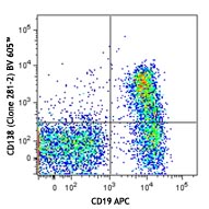
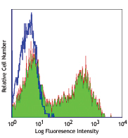
C57BL/6 mouse splenocytes stained with 90 PE/Cyanine7
| Cat # | Size | Price | Quantity Check Availability | Save | ||
|---|---|---|---|---|---|---|
| 102717 | 25 µg | 85€ | ||||
| 102718 | 100 µg | 259€ | ||||
CD38 is a 45 kD type II transmembrane glycoprotein also known as T10. It is an ADP-ribosyl hydrolase expressed at variable levels on hematopoietic cells and in some non-hematopoietic tissues (such as brain, muscle, and kidney). In humans, it is expressed at high levels on plasma cells and activated T and B cells, natural killer (NK) lymphocytes, myeloblasts, and erythroblasts. By functioning as both a cyclase and a hydrolase, CD38 mediates lymphocyte activation, adhesion, and the metabolism of cADPR and NAADP. CD31 is the ligand of CD38.
Product DetailsProduct Details
- Verified Reactivity
- Mouse
- Antibody Type
- Monoclonal
- Host Species
- Rat
- Immunogen
- Mouse bone marrow pre-B cells
- Formulation
- Phosphate-buffered solution, pH 7.2, containing 0.09% sodium azide.
- Preparation
- The antibody was purified by affinity chromatography, and conjugated with PE/Cyanine7 under optimal conditions.
- Concentration
- 0.2 mg/ml
- Storage & Handling
- The antibody solution should be stored undiluted between 2°C and 8°C, and protected from prolonged exposure to light. Do not freeze.
- Application
-
FC - Quality tested
- Recommended Usage
-
Each lot of this antibody is quality control tested by immunofluorescent staining with flow cytometric analysis. For flow cytometric staining, the suggested use of this reagent is ≤ 0.25 µg per 106 cells in 100 µl volume. It is recommended that the reagent be titrated for optimal performance for each application.
- Excitation Laser
-
Blue Laser (488 nm)
Green Laser (532 nm)/Yellow-Green Laser (561 nm)
- Additional Product Notes
- BioLegend is in the process of converting the name PE/Cy7 to PE/Cyanine7. The dye molecule remains the same, so you should expect the same quality and performance from our PE/Cyanine7 products. Please contact Technical Service if you have any questions.
- Product Citations
-
- RRID
-
AB_2072892 (BioLegend Cat. No. 102717)
AB_2275531 (BioLegend Cat. No. 102718)
Antigen Details
- Structure
- ADP-ribosyl hydrolase, 42 kD
- Distribution
-
B cells, NK cells, T cell subset, brain, muscle, kidney
- Function
- Regulation of cellular activation/proliferation, adhesion, metabolism of cADPR and NAADP
- Ligand/Receptor
- CD31, hyaluronic acid
- Antigen References
-
1. Barclay AN, et al. 1997. The Leukocyte Antigen FactsBook Academic Press.
2. Shubinsky G, et al. 1997. Immunity 7:315.
3. Cesano A, et al. 1998. J. Immunol. 160:1106.
4. Cockayne DA, et al. 1998. Blood 92:1324. - Gene ID
- 12494 View all products for this Gene ID
- UniProt
- View information about CD38 on UniProt.org


Other Formats
View All CD38 Reagents Request Custom Conjugation| Description | Clone | Applications |
|---|---|---|
| APC anti-mouse CD38 | 90 | FC |
| FITC anti-mouse CD38 | 90 | FC |
| PE anti-mouse CD38 | 90 | FC |
| Purified anti-mouse CD38 | 90 | FC,IHC-F |
| Alexa Fluor® 488 anti-mouse CD38 | 90 | FC |
| Alexa Fluor® 647 anti-mouse CD38 | 90 | FC,IHC-F |
| PerCP/Cyanine5.5 anti-mouse CD38 | 90 | FC |
| PE/Cyanine7 anti-mouse CD38 | 90 | FC |
| Pacific Blue™ anti-mouse CD38 | 90 | FC |
| Purified anti-mouse CD38 (Maxpar® Ready) | 90 | FC,CyTOF® |
| Alexa Fluor® 594 anti-mouse CD38 | 90 | IHC-F |
| APC/Cyanine7 anti-mouse CD38 | 90 | FC |
| PE/Dazzle™ 594 anti-mouse CD38 | 90 | FC |
| Brilliant Violet 421™ anti-mouse CD38 | 90 | FC |
| TotalSeq™-A0557 anti-mouse CD38 | 90 | PG |
| TotalSeq™-C0557 anti-mouse CD38 | 90 | PG |
| APC/Fire™ 750 anti-mouse CD38 | 90 | FC |
| TotalSeq™-B0557 anti-mouse CD38 | 90 | PG |
| PE/Fire™ 640 anti-mouse CD38 | 90 | FC |
| APC/Fire™ 810 anti-mouse CD38 | 90 | FC |
| Alexa Fluor® 700 anti-mouse CD38 | 90 | FC |
| PE/Fire™ 700 anti-mouse CD38 | 90 | FC |
| Spark YG™ 581 anti-mouse CD38 | 90 | FC |
Customers Also Purchased
Compare Data Across All Formats
This data display is provided for general comparisons between formats.
Your actual data may vary due to variations in samples, target cells, instruments and their settings, staining conditions, and other factors.
If you need assistance with selecting the best format contact our expert technical support team.
-
APC anti-mouse CD38

C57BL/6 mouse splenocytes stained with 90 APC -
FITC anti-mouse CD38

C57BL/6 mouse splenocytes stained with 90 FITC -
PE anti-mouse CD38

C57BL/6 splenocytes stained with 90 FITC -
Purified anti-mouse CD38

C57BL/6 splenocytes stained with purified 90, followed by an... 
Fresh, frozen mouse spleen was stained with purified CD38 cl... -
Alexa Fluor® 488 anti-mouse CD38

C57BL/6 mouse splenocytes stained with 90 Alexa Fluor® 488 -
Alexa Fluor® 647 anti-mouse CD38

C57BL/6 mouse splenocytes stained with 90 Alexa Fluor® 647 
C57BL/6 mouse frozen spleen section was fixed with 4% parafo... -
PerCP/Cyanine5.5 anti-mouse CD38

C57BL/6 mouse splenocytes were stained with CD45R/B220 FITC ... -
PE/Cyanine7 anti-mouse CD38

C57BL/6 mouse splenocytes stained with 90 PE/Cyanine7 -
Pacific Blue™ anti-mouse CD38

C57BL/6 mouse splenocytes stained with 90 Pacific Blue&trade... -
Purified anti-mouse CD38 (Maxpar® Ready)

C57BL/6 mouse splenocytes stained with 152Sm-anti-CD3e (145-... -
Alexa Fluor® 594 anti-mouse CD38

C57BL/6 mouse frozen spleen section was fixed with 4% parafo... -
APC/Cyanine7 anti-mouse CD38

C57BL/6 mouse splenocytes were stained with CD38 (clone 90) ... -
PE/Dazzle™ 594 anti-mouse CD38

C57BL/6 mouse splenocytes were stained with CD38 (clone 90) ... -
Brilliant Violet 421™ anti-mouse CD38

C57BL/6 mouse splenocytes were stained with CD38 (clone 90) ... -
TotalSeq™-A0557 anti-mouse CD38
-
TotalSeq™-C0557 anti-mouse CD38
-
APC/Fire™ 750 anti-mouse CD38

C57BL/6 splenocytes were stained with CD45R/B220 FITC and CD... -
TotalSeq™-B0557 anti-mouse CD38
-
PE/Fire™ 640 anti-mouse CD38

C57BL/6 mouse splenocytes were stained with anti-mouse CD45R... -
APC/Fire™ 810 anti-mouse CD38

C57BL/6 mouse splenocytes were stained with anti-mouse CD45R... -
Alexa Fluor® 700 anti-mouse CD38

C57BL/6 splenocytes were stained with anti-mouse CD45R/B220 ... -
PE/Fire™ 700 anti-mouse CD38

C57BL/6 mouse splenocytes were stained with anti-mouse CD45R... -
Spark YG™ 581 anti-mouse CD38

C57BL/6 splenocytes were stained with anti-mouse/human CD45R...


Follow Us