


- Clone
- G043H7 (See other available formats)
- Regulatory Status
- RUO
- Other Names
- BLR2, CDw197, EBI1, CMKBR7
- Isotype
- Mouse IgG2a, κ
- Ave. Rating
- Submit a Review
- Product Citations
- publications

-

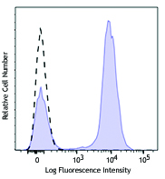
Human peripheral blood lymphocytes were stained with CD3 FITC and CCR7/CD197 (clone G043H7) Brilliant Violet 421™ (top) or mouse IgG2a, κ Brilliant Violet 421™ isotype control (bottom). -

| Cat # | Size | Price | Save |
|---|---|---|---|
| 353207 | 25 tests | ¥47,300 | |
| 353208 | 100 tests | ¥89,540 |
CCR7, also known as CD197, is a chemokine receptor that binds CCL19 and CCL21. CCR7 and its ligands link innate and adaptive immunity by affecting interactions between T cells and dendritic cells and their downstream effect. Naïve T cells enter the lymph node through high endothelial venules, which express CCL21. Dendritic cells and macrophages enter the lymph node through afferent lymphatics. The encounter of T cells and dendritic cells in the T cell zone is CCR7-dependent. In addition, during immunological surveillance, B cells recirculate between B-cell-rich compartments (follicles or B cell zones) in secondary lymphoid organs, surveying for antigen. After antigen binding, B cells move to the boundary of B and T zones to interact with T-helper cells; this B cell migration is directed by CCR7 and its ligands. CCR7-positive cancer cell expression has been associated with lymph node metastasis.
Product DetailsProduct Details
- Reactivity
- Human
- Antibody Type
- Monoclonal
- Host Species
- Mouse
- Immunogen
- CCR7-transfected cells
- Formulation
- Phosphate-buffered solution, pH 7.2, containing 0.09% sodium azide and BSA (origin USA).
- Preparation
- The antibody was purified by affinity chromatography and conjugated with Brilliant Violet 421™ under optimal conditions.
- Concentration
- Lot-specific (to obtain lot-specific concentration and expiration, please enter the lot number in our Certificate of Analysis online tool.)
- Storage & Handling
- The antibody solution should be stored undiluted between 2°C and 8°C, and protected from prolonged exposure to light. Do not freeze.
- Application
-
FC - Quality tested
- Recommended Usage
-
Each lot of this antibody is quality control tested by immunofluorescent staining with flow cytometric analysis. For flow cytometric staining, the suggested use of this reagent is 5 µl per million cells in 100 µl staining volume or 5 µl per 100 µl of whole blood.
Brilliant Violet 421™ excites at 405 nm and emits at 421 nm. The standard bandpass filter 450/50 nm is recommended for detection. Brilliant Violet 421™ is a trademark of Sirigen Group Ltd.
Learn more about Brilliant Violet™.
This product is subject to proprietary rights of Sirigen Inc. and is made and sold under license from Sirigen Inc. The purchase of this product conveys to the buyer a non-transferable right to use the purchased product for research purposes only. This product may not be resold or incorporated in any manner into another product for resale. Any use for therapeutics or diagnostics is strictly prohibited. This product is covered by U.S. Patent(s), pending patent applications and foreign equivalents. - Excitation Laser
-
Violet Laser (405 nm)
- Product Citations
- RRID
-
AB_10915137 (BioLegend Cat. No. 353207)
AB_11203894 (BioLegend Cat. No. 353208)
Antigen Details
- Structure
- Chemokine receptor, G protein-coupled receptors (GPCR), seven transmembrane receptor.
- Distribution
-
T cells, B cells, NK, dendritic cells.
- Function
- The chemokine receptor CCR7 plays a pivotal role in the homing of naïve T cells and regulatory T cells to secondary lymphoid organs, and the migration of dendritic cells into afferent lymphatic vessels.
- Ligand/Receptor
- CCL19 and CCL21.
- Cell Type
- B cells, Dendritic cells, NK cells, T cells
- Biology Area
- Immunology
- Molecular Family
- CD Molecules, Cytokine/Chemokine Receptors, GPCR
- Antigen References
-
1. Yanagihara S, et al. 1998. J. Immunol. 161:3096.
2. Charo IF, et al. 2006. N. Engl. J. Med. 354:610.
3. Reif K, et al. 2002. Nature 416:94.
4. Nakata B, et al. 2008. Oncology 74:69.
5. Brodie T. et al. 2013. Cytometry A. 6: 530-2. PubMed
6. Graves A.J. et al. 2014. Cytometry A. 7: 576–9 PubMed
7. Moncunill G. et al. 2014. Cytometry A. 12: 995-8 PubMed - Gene ID
- 1236 View all products for this Gene ID
- UniProt
- View information about CD197 on UniProt.org
Related Pages & Pathways
Pathways


Related FAQs
- What is the F/P ratio range of our BV421™ format antibody reagents?
-
It is lot-specific. On average it ranges between 2-4.
- Does staining at room temperature or even at 37°C help for checking chemokine receptors expression?
-
Due to continuous recycling of many chemokine receptors, it may be worthwhile to consider staining at room temperature or at 37°C if the staining at lower temperature (which can potentially reduce receptor turnover) is not optimal.
Customers Also Purchased

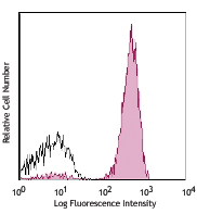
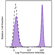
Compare Data Across All Formats
This data display is provided for general comparisons between formats.
Your actual data may vary due to variations in samples, target cells, instruments and their settings, staining conditions, and other factors.
If you need assistance with selecting the best format contact our expert technical support team.


Follow Us