


- Clone
- RMT3-23 (See other available formats)
- Regulatory Status
- RUO
- Other Names
- T cell immunoglobulin and mucin domain containing 3 protein, hepatitis virus cellular receptor 2, CD366
- Isotype
- Rat IgG2a, κ
- Ave. Rating
- Submit a Review
- Product Citations
- publications

-

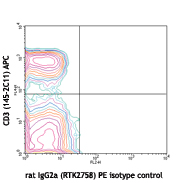
C57BL/6 mouse splenocytes were stained with anti-mouse CD3ε (clone 145-2C11) PerCP/Fire™ 780 and anti-mouse CD366 (Tim-3) (clone RMT3-23) PE (left) or rat IgG2a, κ PE isotype control (right).
| Cat # | Size | Price | Save |
|---|---|---|---|
| 119703 | 50 µg | ¥29,480 | |
| 119704 | 200 µg | ¥89,760 |
CD366 (Tim-3) is a transmembrane protein also known as T cell immunoglobulin and mucin domain containing protein-3. Tim-3 is expressed at high levels on Th1 lymphocytes and CD11b+ macrophages. Tim-3 has also been shown to exist as a soluble protein. Cells expressing Tim-3 are present at high levels in the CNS of animals at the onset of experimental autoimmune encephalomyelitis (EAE), a disease mediated by lymphocytes secreting Th1-like cytokines. Tim-3 has been proposed to inhibit Th1-mediated immune responses and promote immunological tolerance.
Product DetailsProduct Details
- Reactivity
- Mouse
- Antibody Type
- Monoclonal
- Host Species
- Rat
- Formulation
- Phosphate-buffered solution, pH 7.2, containing 0.09% sodium azide.
- Preparation
- The antibody was purified by affinity chromatography, and conjugated with PE under optimal conditions.
- Concentration
- 0.2 mg/ml
- Storage & Handling
- The antibody solution should be stored undiluted between 2°C and 8°C, and protected from prolonged exposure to light. Do not freeze.
- Application
-
FC - Quality tested
- Recommended Usage
-
Each lot of this antibody is quality control tested by immunofluorescent staining with flow cytometric analysis. For flow cytometric staining, the suggested use of this reagent is ≤0.25 µg per million cells in 100 µl volume. It is recommended that the reagent be titrated for optimal performance for each application.
- Excitation Laser
-
Blue Laser (488 nm)
Green Laser (532 nm)/Yellow-Green Laser (561 nm)
- Application Notes
-
Additional reported applications (for relevant formats) include: in vitro1 and in vivo2 blocking of Tim-3, and immunohistochemical staining of frozen sections2. The Ultra-LEAF™ purified antibody (Endotoxin <0.01 EU/µg, Azide-Free, 0.2 µm filtered) is recommended for functional assays (Cat. Nos. 119731-119736).
-
Application References
(PubMed link indicates BioLegend citation) -
- Nakae S, et al. 2007. Blood 110(7):2565-8. (FC, Block)
- Oikawa T, et al. 2006. J. Immunol. 177(7):4281-7. (FC, Block, IHC)
- Product Citations
- RRID
-
AB_345377 (BioLegend Cat. No. 119703)
AB_345378 (BioLegend Cat. No. 119704)
Antigen Details
- Structure
- Transmembrane protein containing immunoglobulin domain and mucin-like domain; predicted molecular weight 31 kD; can exist as a soluble form lacking mucin and transmembrane domains
- Distribution
-
Expressed on activated Th1 lymphocytes and CD11b+ macrophages
- Function
- May play a role in the development of immune responses and the development of Th1-mediated responses
- Ligand/Receptor
- Putative ligand on resting CD4+ lymphocytes
- Cell Type
- Macrophages, Th1
- Biology Area
- Immunology, Inhibitory Molecules
- Molecular Family
- CD Molecules, Immune Checkpoint Receptors
- Antigen References
-
1. Sabatos CA, et al. 2003. Nat. Immunol. 4:1102.
2. Sanchez-Fueyo A, et al. 2003. Nat. Immunol. 4:1102.
3. Kuchroo VK, et al. 2003. Nat. Rev. Immunol. 3:454.
4. Mooney L, et al. 2002. Nature 415:536. - Gene ID
- 171285 View all products for this Gene ID
- UniProt
- View information about CD366 on UniProt.org


Related FAQs
- What type of PE do you use in your conjugates?
- We use R-PE in our conjugates.
Customers Also Purchased
Compare Data Across All Formats
This data display is provided for general comparisons between formats.
Your actual data may vary due to variations in samples, target cells, instruments and their settings, staining conditions, and other factors.
If you need assistance with selecting the best format contact our expert technical support team.


Follow Us