


- Clone
- 67A4 (See other available formats)
- Regulatory Status
- RUO
- Other Names
- E-Cadherin, cadherin-1, and UVO
- Isotype
- Mouse IgG1, κ
- Ave. Rating
- Submit a Review
- Product Citations
- publications

-

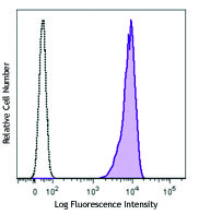
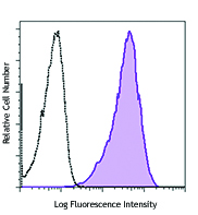
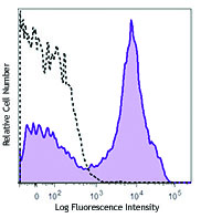
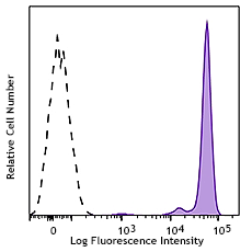
Human colon carcinoma cell line (HT29) was stained with CD324 (clone 67A4) PE/Cyanine7 (filled histogram) or mouse IgG1, κ PE/Cyanine7 isotype control (open histogram).
| Cat # | Size | Price | Save |
|---|---|---|---|
| 324115 | 25 tests | ¥38,720 | |
| 324116 | 100 tests | ¥84,040 |
The 67A4 antibody recognizes human CD324 also known as E-cadherin, cadherin-1, and UVO. CD324, a member of the cadherin superfamily, is a calcium-dependent, transmembrane cell-cell adhesion glycoprotein composed of 4 extracellular cadherin repeats and a highly conserved cytoplasmic tail region with a predicted molecular weight of approximately 100 kD. CD324 is widely expressed in epithelial cells in the colon, uterus, liver, keratinocytes, brain, heart, muscle, kidney, and pancreas, as well as erythroid cells. CD324 functions as a cell adhesion molecule involved in development, bacterial pathogenesis, and tumor invasion. In bacterial pathogenesis, the ectodomain of CD324 mediates bacterial adhesion to mammalian cells, while the cytoplasmic domain is required for internalization. CD324 binds to the αEβ7 integrin to mediate cell adhesion and also interacts with a number of intracellular proteins including including erbin, ezrin, caspase-3, caspase 8, β-catenin, presenilin 1, casein kinase II , as well as other extracellular proteins including the EGF receptor. CD324 is phosphorylated on multiple residues (S857, S866, S870, S872), and can be proteolytically cleaved at reside D769 by caspase-3. The 67A4 antibody has been shown to be useful for flow cytometry.
Product DetailsProduct Details
- Reactivity
- Human
- Antibody Type
- Monoclonal
- Host Species
- Mouse
- Immunogen
- T-47D cells
- Formulation
- Phosphate-buffered solution, pH 7.2, containing 0.09% sodium azide and BSA (origin USA)
- Preparation
- The antibody was purified by affinity chromatography and conjugated with PE/Cyanine7 under optimal conditions.
- Concentration
- Lot-specific (to obtain lot-specific concentration and expiration, please enter the lot number in our Certificate of Analysis online tool.)
- Storage & Handling
- The antibody solution should be stored undiluted between 2°C and 8°C, and protected from prolonged exposure to light. Do not freeze.
- Application
-
FC - Quality tested
- Recommended Usage
-
Each lot of this antibody is quality control tested by immunofluorescent staining with flow cytometric analysis. For flow cytometric staining, the suggested use of this reagent is 5 µl per million cells in 100 µl staining volume or 5 µl per 100 µl of whole blood.
It is also recommended using EDTA-based solutions for dissociating attachment-dependent cell lines. - Excitation Laser
-
Blue Laser (488 nm)
Green Laser (532 nm)/Yellow-Green Laser (561 nm)
- Additional Product Notes
-
BioLegend is in the process of converting the name PE/Cy7 to PE/Cyanine7. The dye molecule remains the same, so you should expect the same quality and performance from our PE/Cyanine7 products. Please contact Technical Service if you have any questions.
-
Application References
(PubMed link indicates BioLegend citation) -
- Armeanu S, et al. 1995. J. Cell Biol. 131:243.
- Bühring HJ, et al. 1996. Leukemia 10:106.
- Yauch RL, et al. 2005. Clin. Cancer Res. 11:8686. (WB)
- Oeztuerk-Winder F, et al. 2012. EMBO J. 31:3431. (FC) PubMed
- Ardehali R, et al. 2013. PNAS. 110:3405. PubMed
- Rasanen K, et al. 2013. Mol Cell Protecomics. 12:3778. PubMed
- Chaudhury A, et al. 2014. Nucleic Acids Res. 42:86. PubMed
- Milne P, et al. 2015. Blood. 125:470. PubMed
- Product Citations
- RRID
-
AB_2563095 (BioLegend Cat. No. 324115)
AB_2563096 (BioLegend Cat. No. 324116)
Antigen Details
- Structure
- Member of the cadherin superfamily. Calcium-dependent, transmembrane cell-cell adhesion glycoprotein composed of 4 extracellular cadherin repeats and a highly conserved cytoplasmic tail region. Predicted molecular weight approximately 100 kD.
- Distribution
-
Widely expressed in epithelial cells in the colon, uterus, liver, keratinocytes, brain, heart, muscle, kidney, and pancreas, as well as erythroid cells
- Function
- Cell adhesion molecule involved in development, bacterial pathogenesis, and tumor invasion. The ectodomain of CD324 mediates bacterial adhesion to mammalian cell, while the cytoplasmic domain is required for internalization.
- Interaction
- Interacts with a variety of proteins including erbin, ezrin, caspase-3, caspase 8, EGF receptor, β-catenin, presenilin 1, casein kinase II, and others
- Ligand/Receptor
- αEβ7 integrin
- Modification
- Phosphorylated on multiple residues (S857, S866, S870, S872), proteolytically cleaved at reside D769 by caspase-3.
- Cell Type
- Embryonic Stem Cells, Epithelial cells, Erythrocytes
- Biology Area
- Cell Biology, Immunology, Neuroscience, Stem Cells, Synaptic Biology
- Molecular Family
- Adhesion Molecules, CD Molecules
- Antigen References
-
1. Overduin M, et al. 1995. Science 267:386.
2. Boggon TJ, et al. 2002. Science 296:1303.
3. Berx G, et al. 1995. EMBO J. 14:6107.
4. Perl AK, et al. 1998. Nature 392:190. - Gene ID
- 999 View all products for this Gene ID
- UniProt
- View information about CD324 on UniProt.org
Related Pages & Pathways
Pathways


Related FAQs
Customers Also Purchased
Compare Data Across All Formats
This data display is provided for general comparisons between formats.
Your actual data may vary due to variations in samples, target cells, instruments and their settings, staining conditions, and other factors.
If you need assistance with selecting the best format contact our expert technical support team.


Follow Us