 Login / Register


- Clone
- AK4 (See other available formats)
- Regulatory Status
- RUO
- Workshop
- VI P-44
- Other Names
- GMP-140, PADGEM
- Isotype
- Mouse IgG1, κ
- Ave. Rating
- Submit a Review
- Product Citations
- publications

-

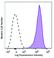
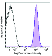
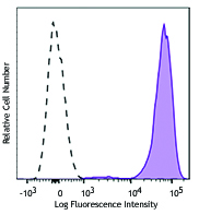
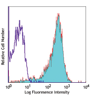
Thrombin-activated human peripheral blood platelets were stained with CD62P (P-Selectin) (clone AK4) Brilliant Violet 785™ (filled histogram) or mouse IgG1, κ Brilliant Violet 785™ isotype control (open histogram).
| Cat # | Size | Price | Quantity Check Availability | Save | ||
|---|---|---|---|---|---|---|
| 304941 | 25 tests | 168€ | ||||
| 304942 | 100 tests | 340€ | ||||
CD62P is a 140 kD type I transmembrane glycoprotein also known as P-selectin, platelet activation-dependent granule membrane protein (PADGEM), and GMP-140. It is expressed on activated platelets, megakaryocytes, and endothelial cells. CD62P is primarily stored in secretory α-granules in platelets and Weibel-Palade bodies in endothelial cells, and is rapidly relocated to the plasma membrane upon activation. The ligands for CD62P are CD162 and CD24. A primary function of CD62P is cell adhesion during neutrophil rolling, and platelet-neutrophil and platelet-monocyte interactions.
Product DetailsProduct Details
- Reactivity
- Human
- Antibody Type
- Monoclonal
- Host Species
- Mouse
- Formulation
- Phosphate-buffered solution, pH 7.2, containing 0.09% sodium azide and BSA (origin USA).
- Preparation
- The antibody was purified by affinity chromatography and conjugated with Brilliant Violet 785™ under optimal conditions.
- Concentration
- Lot-specific (to obtain lot-specific concentration and expiration, please enter the lot number in our Certificate of Analysis online tool.)
- Storage & Handling
- The antibody solution should be stored undiluted between 2°C and 8°C, and protected from prolonged exposure to light. Do not freeze.
- Application
-
FC - Quality tested
- Recommended Usage
-
Each lot of this antibody is quality control tested by immunofluorescent staining with flow cytometric analysis. For flow cytometric staining, the suggested use of this reagent is 5 µl per million cells in 100 µl staining volume or 5 µl per 100 µl of whole blood.
Brilliant Violet 785™ excites at 405 nm and emits at 785 nm. The bandpass filter 780/60 nm is recommended for detection, although filter optimization may be required depending on other fluorophores used. Be sure to verify that your cytometer configuration and software setup are appropriate for detecting this channel. Refer to your instrument manual or manufacturer for support. Brilliant Violet 785™ is a trademark of Sirigen Group Ltd.
Learn more about Brilliant Violet™.
This product is subject to proprietary rights of Sirigen Inc. and is made and sold under license from Sirigen Inc. The purchase of this product conveys to the buyer a non-transferable right to use the purchased product for research purposes only. This product may not be resold or incorporated in any manner into another product for resale. Any use for therapeutics or diagnostics is strictly prohibited. This product is covered by U.S. Patent(s), pending patent applications and foreign equivalents. - Excitation Laser
-
Violet Laser (405 nm)
- Application Notes
-
Additional reported applications (for the relevant formats) include: immunohistochemical staining of acetone-fixed frozen tissue sections4 and in vitro blocking of adhesion of platelets1. The Ultra-LEAF™ purified antibody (Endotoxin < 0.01 EU/µg, Azide-Free, 0.2 µm filtered) is recommended for functional assays (Cat. No. 304947 & 304948).
- Application References
-
- Skinner M, et al. 1991. J. Biol. Chem. 266:5371. (Block)
- Kishimoto T, et al. Eds. 1997. Leucocyte Typing VI. Garland Publishing Inc. London.
- Yen YT, et al. 2006. J. Virol. 80:2684.
- Sato Y, et al. 2005. Blood 106:428. (IHC)
- Product Citations
- RRID
-
AB_2810451 (BioLegend Cat. No. 304941)
AB_2810452 (BioLegend Cat. No. 304942)
Antigen Details
- Structure
- Selectin, type I glycoprotein, 140 kD
- Distribution
-
Activated platelets, megakaryocytes, endothelial cells
- Function
- Adhesion, neutrophil rolling, platelet-neutrophil and platelet-monocyte interactions
- Ligand/Receptor
- CD162 (PSGL-1), CD24 and sialylated Lewis X
- Cell Type
- Endothelial cells, Megakaryocytes, Neutrophils, Platelets
- Biology Area
- Cell Adhesion, Cell Biology, Immunology, Neuroscience, Synaptic Biology
- Molecular Family
- Adhesion Molecules, CD Molecules
- Antigen References
-
1. McEver R, et al. 1995. J. Biol. Chem. 270:11025.
2. Varki A. 1994. P. Natl. Acad. Sci. USA 91:7390. - Gene ID
- 6403 View all products for this Gene ID
- UniProt
- View information about CD62P on UniProt.org
Related Pages & Pathways
Pathways


Customers Also Purchased
Compare Data Across All Formats
This data display is provided for general comparisons between formats.
Your actual data may vary due to variations in samples, target cells, instruments and their settings, staining conditions, and other factors.
If you need assistance with selecting the best format contact our expert technical support team.


Follow Us