 Login / Register


- Clone
- 6D5 (See other available formats)
- Regulatory Status
- RUO
- Other Names
- B4
- Isotype
- Rat IgG2a, κ
- Ave. Rating
- Submit a Review
- Product Citations
- publications

-

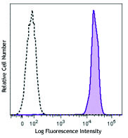
C57BL/6 mouse splenocytes were stained with CD19 (clone 6D5) Brilliant Violet 785™.
| Cat # | Size | Price | Quantity Check Availability | Save | ||
|---|---|---|---|---|---|---|
| 115543 | 125 µL | 168€ | ||||
CD19 is a 95 kD glycoprotein also known as B4. It is a member of the Ig superfamily, expressed on all pro-B to mature B cells (during development) and follicular dendritic cells. Plasma cells do not express CD19. CD19, in association with CD21 and CD81, forms a molecular complex integral to B cell activation.
Product DetailsProduct Details
- Reactivity
- Mouse
- Antibody Type
- Monoclonal
- Host Species
- Rat
- Immunogen
- Mouse CD19-expressing K562 human erythroleukemia cells
- Formulation
- Phosphate-buffered solution, pH 7.2, containing 0.09% sodium azide and BSA (origin USA).
- Preparation
- The antibody was purified by affinity chromatography and conjugated with Brilliant Violet 785™ under optimal conditions.
- Concentration
- Lot-specific (to obtain lot-specific concentration and expiration, please enter the lot number in our Certificate of Analysis online tool.)
- Storage & Handling
- The antibody solution should be stored undiluted between 2°C and 8°C, and protected from prolonged exposure to light. Do not freeze.
- Application
-
FC - Quality tested
- Recommended Usage
-
Each lot of this antibody is quality control tested by immunofluorescent staining with flow cytometric analysis. For flow cytometric staining, the suggested use of this reagent is 5 µl per million cells in 100 µl staining volume or 5 µl per 100 µl of whole blood. It is recommended that the reagent be titrated for optimal performance for each application.
Brilliant Violet 785™ excites at 405 nm and emits at 785 nm. The bandpass filter 780/60 nm is recommended for detection, although filter optimization may be required depending on other fluorophores used. Be sure to verify that your cytometer configuration and software setup are appropriate for detecting this channel. Refer to your instrument manual or manufacturer for support. Brilliant Violet 785™ is a trademark of Sirigen Group Ltd.
Learn more about Brilliant Violet™.
This product is subject to proprietary rights of Sirigen Inc. and is made and sold under license from Sirigen Inc. The purchase of this product conveys to the buyer a non-transferable right to use the purchased product for research purposes only. This product may not be resold or incorporated in any manner into another product for resale. Any use for therapeutics or diagnostics is strictly prohibited. This product is covered by U.S. Patent(s), pending patent applications and foreign equivalents. - Excitation Laser
-
Violet Laser (405 nm)
- Application Notes
-
Additional reported applications (for the relevant formats) include: immunofluorescence7.
- Application References
-
- Shoham T, et al. 2003. J. Immunol. 171:4062. (FC)
- Goodyear CS, et al. 2004. J. Immunol. 172:2870. (FC)
- Kamimura D, et al. 2006. J. Immunol. 177:306. (FC)
- Andoniou CE, et al. 2005. Nat. Immunol. 6:1011. (FC)
- Lawson BR, et al. 2007. J. Immunol. 178:5366. (FC)
- Phan TG, et al. 2007. Nat. Immunol. 8:992. (FC)
- Hayashida K, et al. 2008. J. Biol. Chem. 283:19895. (IF) PubMed
- Charles N, et al. 2010. Nat. Med. 16:701. (FC) PubMed
- Bankoti J, et al. 2010. Toxicol. Sci. 115:422. (FC) PubMed
- Stadnisky MD, et al. 2011. Blood. 117:5133. (FC) PubMed
- Perlot T, et al. 2012. J. Immunol. 188:1201. (FC) PubMed
- Olive V, et al. 2013. Elife. 2:822. PubMed
- Miyai T, et al. 2014. PNAS. 111:11780. PubMed
- Product Citations
- RRID
-
AB_11218994 (BioLegend Cat. No. 115543)
Antigen Details
- Structure
- Ig superfamily, associates with CD21 and CD81, 95 kD
- Distribution
-
Pro-B cells to mature B cells (during development), follicular dendritic cells
- Function
- Modulates B cell activation and differentiation
- Ligand/Receptor
- CD21, CD81, Leu-13
- Cell Type
- B cells, Dendritic cells
- Biology Area
- Costimulatory Molecules, Immunology
- Molecular Family
- CD Molecules
- Antigen References
-
1. Fearon DT. 1993. Curr. Opin. Immunol. 5:341.
2. Krop I, et al. 1996. Eur. J. Immunol. 26:238.
3. Krop I, et al. 1996. J. Immunol. 157:48.
4. Tedder TF, et al. 1994. Immunol. Today 15:437. - Gene ID
- 12478 View all products for this Gene ID
- UniProt
- View information about CD19 on UniProt.org
Related Pages & Pathways
Pathways


Related FAQs
Customers Also Purchased
Compare Data Across All Formats
This data display is provided for general comparisons between formats.
Your actual data may vary due to variations in samples, target cells, instruments and their settings, staining conditions, and other factors.
If you need assistance with selecting the best format contact our expert technical support team.


Follow Us