 Login / Register


- Clone
- P44-8 (See other available formats)
- Regulatory Status
- RUO
- Other Names
- NKp44, NCR2, Activating NK receptor NKp44, natural cytotoxicity triggering receptor 2. lymphocyte antigen 95 homolog
- Isotype
- Mouse IgG1, κ
- Ave. Rating
- Submit a Review
- Product Citations
- publications

-

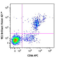
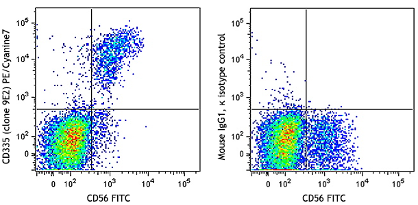
Human peripheral blood lymphocytes were stimulated with recombinant human IL-2 for 7 days and then stained with CD56 APC and CD336 (clone P44-8) PE/Cyanine7 (left) or mouse IgG1, κ PE/Cyanine7 isotype control (right).
| Cat # | Size | Price | Quantity Check Availability | Save | ||
|---|---|---|---|---|---|---|
| 325115 | 25 tests | 128€ | ||||
| 325116 | 100 tests | 306€ | ||||
CD336 is also known as activating NK receptor NKp44 (NKp44), natural cytotoxicity triggering receptor 2, and lymphocyte antigen 95 homolog. It is a type I transmembrane protein, member of the natural cytotoxicity receptor family that contains one immunoglobulin-like domain. NKp44 has an apparent molecular weight of 44 kD and three isoforms are produced by alternative splicing. NKp44 is expressed on IL-2 activated NK cells and a subset of γ/δ T cells. NKp44 enhances NK cell mediated cytolysis of virus infected cells and tumor cells. NKp44 has been shown to associate with the intracellular adaptor DAP12.
Product DetailsProduct Details
- Reactivity
- Human
- Antibody Type
- Monoclonal
- Host Species
- Mouse
- Immunogen
- recombinant human NKp44
- Formulation
- Phosphate-buffered solution, pH 7.2, containing 0.09% sodium azide and BSA (origin USA)
- Preparation
- The antibody was purified by affinity chromatography and conjugated with PE/Cyanine7 under optimal conditions.
- Concentration
- Lot-specific (to obtain lot-specific concentration and expiration, please enter the lot number in our Certificate of Analysis online tool.)
- Storage & Handling
- The antibody solution should be stored undiluted between 2°C and 8°C, and protected from prolonged exposure to light. Do not freeze.
- Application
-
FC - Quality tested
- Recommended Usage
-
Each lot of this antibody is quality control tested by immunofluorescent staining with flow cytometric analysis. For flow cytometric staining, the suggested use of this reagent is 5 µl per million cells in 100 µl staining volume or 5 µl per 100 µl of whole blood.
- Excitation Laser
-
Blue Laser (488 nm)
Green Laser (532 nm)/Yellow-Green Laser (561 nm)
- Application Notes
-
The p44-8 antibody against human NKp44 has been shown to be useful for flow cytometry, stimulation of human NK cells via NKp44 in a redirected lysis assay, and blocking of NKp44 function in solution. Additional reported applications (for the relevant formats) include: stimulation of human NK cells via NKp44 in a redirected lysis assay, and blocking of NKp44 function in solution1,2. The LEAF™ purified antibody (Endotoxin < 0.1 EU/µg, Azide-Free, 0.2 µm filtered) is recommended for functional assays (Cat. No. 325104).
- Additional Product Notes
-
BioLegend is in the process of converting the name PE/Cy7 to PE/Cyanine7. The dye molecule remains the same, so you should expect the same quality and performance from our PE/Cyanine7 products. Please contact Technical Service if you have any questions.
- Application References
- Product Citations
- RRID
-
AB_2616753 (BioLegend Cat. No. 325115)
AB_2616754 (BioLegend Cat. No. 325116)
Antigen Details
- Structure
- Type I transmembrane protein, member of the natural cytotoxicity receptor family, contains one immunoglobulin-like domain. Approximately 44 kD. Three isoforms produced by alternative splicing.
- Distribution
-
IL-2 activated NK cells and cell lines, some γ/δ T cells activated in vitro
- Function
- NK cell triggering of cytolysis toward tumor targets and other target cells deficient in MHC class I molecules
- Interaction
- DAP12
- Cell Type
- NK cells, T cells
- Biology Area
- Immunology
- Molecular Family
- CD Molecules
- Antigen References
-
1. Cantoni C, et al. 1999. J. Exp. Med. 189:787.
2. Allcock RJN, et al. 2003. Eur. J. Immunol. 33:567.
3. Cantoni C, et al. 2003. Structure. 11:725. - Gene ID
- 9436 View all products for this Gene ID
- UniProt
- View information about CD336 on UniProt.org
Related Pages & Pathways
Pathways


Customers Also Purchased
Compare Data Across All Formats
This data display is provided for general comparisons between formats.
Your actual data may vary due to variations in samples, target cells, instruments and their settings, staining conditions, and other factors.
If you need assistance with selecting the best format contact our expert technical support team.


Follow Us