 Login / Register


- Clone
- 3G8 (See other available formats)
- Regulatory Status
- RUO
- Workshop
- V NK80
- Other Names
- FcγRIII, Fc gamma receptor, Fc gamma receptor 3
- Isotype
- Mouse IgG1, κ
- Ave. Rating
- Submit a Review
- Product Citations
- publications

-

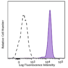
Human peripheral blood lymphocytes stained with 3G8 PerCP
| Cat # | Size | Price | Quantity Check Availability | Save | ||
|---|---|---|---|---|---|---|
| 302029 | 25 tests | 123€ | ||||
| 302030 | 100 tests | 268€ | ||||
CD16 is known as low affinity IgG receptor III (FcγRIII). It is expressed as two distinct forms (CD16a and CD16b). CD16a (FcγRIIIA) is a 50-65 kD polypeptide-anchored transmembrane protein. It is expressed on the surface of NK cells, activated monocytes, macrophages, and placental trophoblasts in humans. CD16b (FcγRIIIB) is a 48 kD glycosylphosphatidylinositol (GPI)-anchored protein. Its extracellular domain is over 95% homologous to that of CD16a, and it is expressed specifically on neutrophils. CD16 binds aggregated IgG or IgG-antigen complex which functions in NK cell activation, phagocytosis, and antibody-dependent cell-mediated cytotoxicity (ADCC).
Product DetailsProduct Details
- Reactivity
- Human,Cynomolgus,Rhesus
- Antibody Type
- Monoclonal
- Host Species
- Mouse
- Immunogen
- Human PMN cells
- Formulation
- Phosphate-buffered solution, pH 7.2, containing 0.09% sodium azide and BSA (origin USA)
- Preparation
- The antibody was purified by affinity chromatography, and conjugated with PerCP under optimal conditions.
- Concentration
- Lot-specific (to obtain lot-specific concentration and expiration, please enter the lot number in our Certificate of Analysis online tool.)
- Storage & Handling
- The antibody solution should be stored undiluted between 2°C and 8°C, and protected from prolonged exposure to light. Do not freeze.
- Application
-
FC - Quality tested
- Recommended Usage
-
Each lot of this antibody is quality control tested by immunofluorescent staining with flow cytometric analysis. For flow cytometric staining, the suggested use of this reagent is 5 µl per million cells in 100 µl staining volume or 5 µl per 100 µl of whole blood.
* PerCP has a maximum absorption of 482 nm and a maximum emission of 675 nm. - Excitation Laser
-
Blue Laser (488 nm)
- Application Notes
-
The 3G8 antibody clone blocks neutrophil phagocytosis and stimulates NK cell proliferation. It has been reported that this clone interacts with the FcγRIIa and FcγRIIIb receptors causing neutrophil activation and aggregation18. Due to this phenomenon staining in whole blood may cause a reduction in the number of granulocytes or alter their scatter profile.
Additional reported applications (for the relevant formats) include: immunohistochemical staining of acetone-fixed frozen tissue sections6, immunoprecipitation3, stimulation of NK cell proliferation4, blocking of phagocytosis5, and blocking of immunoglobulin binding to FcγRIII7,8. The Ultra-LEAF™ purified antibody (Endotoxin < 0.01 EU/µg, Azide-Free, 0.2 µm filtered) is recommended for functional assays (Cat. No. 302049, 302050, 302057, 302058). - Application References
-
- Knapp W, et al. Eds. 1989. Leucocyte Typing IV. Oxford University Press. New York.
- Schlossman S, et al. Eds. 1995. Leucocyte Typing V. Oxford University Press. New York.
- Edberg J, et al. 1997. J. Immunol. 159:3849. (IP)
- Hoshino S, et al. 1991. Blood 78:3232. (Stim)
- Tamm A, et al. 1996. Immunol. 157:1576. (Block)
- Da Silva DM, et al. 2001. Int. Immunol. 13:633. (IHC)
- Holl V, et al. 2004. J. Immunol. 173:6274. (Block)
- Hober D, et al. 2002. J. Gen. Virol. 83:2169. (Block)
- Brainard DM, et al. 2009. J. Virol. 83:7305. PubMed
- Smed-Sörensen A, et al. 2008. Blood 111:5037. (Block) PubMed
- Timmerman KL, et al. 2008. J. Leukoc. Biol. 84:1271. (FC) PubMed
- Yoshino N, et al. 2000. Exp. Anim. (Tokyo) 49:97. (FC)
- Rout N, et al. 2010. PLoS One 5:e9787. (FC)
- Kim WK, et al. 2006. Am. J. Pathol. 168:822. (FC)
- Boltz A, et al. 2011. J. Biol Chem. 286:21896. PubMed
- Wu Z, et al. 2013. J. Virol. 87:7717. PubMed
- Peterson VM, et al. 2017. Nat. Biotechnol. 35:936. (PG)
- Vossebeld PJ, et al. 1997. Biochem J. 323:87-94 (Stim)
- Product Citations
- RRID
-
AB_940378 (BioLegend Cat. No. 302029)
AB_940380 (BioLegend Cat. No. 302030)
Antigen Details
- Structure
- Ig superfamily, transmembrane form (50-65 kD) or GPI-linked form (48 kD)
- Distribution
-
NK cells, activated monocytes, macrophages, neutrophils
- Function
- Low affinity IgG Fc receptor, phagocytosis, ADCC
- Ligand/Receptor
- Aggregated IgG, IgG-antigen complex
- Cell Type
- Dendritic cells, Macrophages, Monocytes, Neutrophils, NK cells
- Biology Area
- Immunology, Innate Immunity
- Molecular Family
- CD Molecules, Fc Receptors
- Antigen References
-
1. Fleit H, et al. 1982. P. Natl. Acad. Sci. USA 79:3275.
2. Stroncek D, et al. 1991. Blood 77:1572.
3. Wirthmueller U, et al. 1992. J. Exp. Med. 175:1381. - Gene ID
- 2214 View all products for this Gene ID
- UniProt
- View information about CD16 on UniProt.org
Related Pages & Pathways
Pathways


Related FAQs
- Is our human Trustain FcX™ (cat# 422302) compatible with anti human CD16, CD32 and CD64 clones 3G8, FUN-2 and 10.1 respectively?
-
Yes
- How stable is PerCP/Cy5.5 tandem as compared to PerCP alone?
-
PerCP/Cy5.5 is quite photostable and also better than PerCP alone in withstanding fixation.
Customers Also Purchased
Compare Data Across All Formats
This data display is provided for general comparisons between formats.
Your actual data may vary due to variations in samples, target cells, instruments and their settings, staining conditions, and other factors.
If you need assistance with selecting the best format contact our expert technical support team.


Follow Us