 Login / Register


- Clone
- 5A6 (See other available formats)
- Regulatory Status
- RUO
- Other Names
- S5.7, CVID6, TSPAN28
- Isotype
- Mouse IgG1, κ
- Ave. Rating
- Submit a Review
- Product Citations
- publications

-

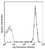
Human peripheral blood lymphocytes stained with purified CD81 (5A6) conjugated with PE
| Cat # | Size | Price | Quantity Check Availability | Save | ||
|---|---|---|---|---|---|---|
| 349501 | 25 µg | 44€ | ||||
| 349502 | 100 µg | 109€ | ||||
CD81 is a 26 kD non-glycosylated member of the tetraspanin superfamily (TM4SF), also known as TAPA-1 (target of an antiproliferative antibody). CD81 is expressed on T and B cells, NK cells, monocytes, dendritic cells, thymocytes, endothelial cells, and fibroblasts. It also has low levels of expression on granulocytes. CD81 induces B cell adhesion via VLA-4 integrin and has been shown to play a role in early T cell development. CD81 associates with several other cell-surface proteins in a multimolecular complex, including CD19, CD21, CD20, CD37, CD53, and CD82 in B cells, and CD4, CD8, and CD82 in T cells.
Product DetailsProduct Details
- Reactivity
- Human
- Antibody Type
- Monoclonal
- Host Species
- Mouse
- Immunogen
- Human OCI-LY8 cell line
- Formulation
- Phosphate-buffered solution, pH 7.2, containing 0.09% sodium azide.
- Preparation
- The antibody was purified by affinity chromatography.
- Concentration
- 0.5 mg/ml
- Storage & Handling
- The antibody solution should be stored undiluted between 2°C and 8°C.
- Application
-
FC - Quality tested
WB, IP - Reported in the literature, not verified in house - Recommended Usage
-
Each lot of this antibody is quality control tested by immunofluorescent staining with flow cytometric analysis. For flow cytometric staining, the suggested use of this reagent is ≤0.5 µg per million cells in 100 µl volume. It is recommended that the reagent be titrated for optimal performance for each application.
- Application Notes
-
Additional reported applications (for the relevant formats) include: Western Blotting3 and immunoprecipitation2,3.
- Application References
-
- Van Zelm MC, et al. 2010. J. Clin. Invest. 120:1265.
- Oren R, et al. 1990. Mol. Cell. Biol. 8:4007. (IP)
- Clark K, et al. 2004. J. Biol. Chem. 279(19):19401. (IP, WB)
- Mochida K, et al. 2008. J. Virol. 13:6711.
- Rappa G, et al. 2014. Mol Cancer Res. 12:1840. PubMed
- Product Citations
- RRID
-
AB_10642023 (BioLegend Cat. No. 349501)
AB_10643417 (BioLegend Cat. No. 349502)
Antigen Details
- Structure
- 26 kD, type III transmembrane protein, member of the TM4SF tetraspanin family. Complexed with CD82, CD19, CD21, or CD4, CD8.
- Distribution
-
T cells, NK, monocytes, B cells, endothelial and epithelial cells, low on granulocytes.
- Function
- Regulates cell activation and growth and cell aggregation.
- Cell Type
- B cells, Endothelial cells, Epithelial cells, Monocytes, Neural Stem Cells, NK cells, T cells
- Biology Area
- Cell Biology, Immunology, Neuroscience, Neuroscience Cell Markers, Signal Transduction, Stem Cells, Transcription Factors
- Molecular Family
- Adhesion Molecules, CD Molecules
- Antigen References
-
- Van Zelm MC, et al. 2010. J. Clin. Invest. 120:1265.
- Fearon D, et al. 1995. Annu. Rev. Immunol. 13:127.
- Wright M, et al. 1994. Immunol. Today 15:588.
- Gene ID
- 975 View all products for this Gene ID
- UniProt
- View information about CD81 on UniProt.org


Customers Also Purchased
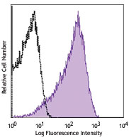

Compare Data Across All Formats
This data display is provided for general comparisons between formats.
Your actual data may vary due to variations in samples, target cells, instruments and their settings, staining conditions, and other factors.
If you need assistance with selecting the best format contact our expert technical support team.


Follow Us