 Login / Register


- Clone
- CD28.2 (See other available formats)
- Regulatory Status
- RUO
- Workshop
- V-CD28.05
- Other Names
- T44, Tp44
- Isotype
- Mouse IgG1, κ
- Ave. Rating
- Submit a Review
- Product Citations
- publications

-

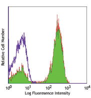
Human peripheral blood lymphocytes stained with LEAF™ purified CD28.2, followed by anti-mouse IgGs FITC -

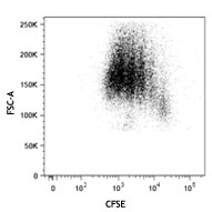
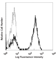
Human peripheral blood mononuclear cells were stained with CFSE on day 0, and then stimulated with (filled histogram) or without (open histogram) immobilized LEAF™ Purified CD3 (clone UCHT1) and LEAF™ purified CD28 (clone CD28.2) for 3 days. On day 4, cells were harvested and stained with CD4 Brilliant Violet 711™. Dot plot (above) was analyzed on live cells. Histogram data (below) was analyzed by gating on CD4 positive cells (above). -

Human peripheral blood mononuclear cells were stained with CFSE on day 0, and then stimulated with (filled histogram) or without (open histogram) immobilized LEAF™ Purified CD3 (clone UCHT1) and LEAF™ purified CD28 (clone CD28.2) for 3 days. On day 4, cells were harvested and stained with CD4 Brilliant Violet 711™. Dot plot (above) was analyzed on live cells. Histogram data (below) was analyzed by gating on CD4 positive cells (above).
Select size of product is eligible for a 40% discount! Promotion valid until September 30, 2024. Exclusions apply. To view full promotion terms and conditions or to contact your local BioLegend representative to receive a quote, visit our webpage.
CD28 is a 44 kD disulfide-linked homodimeric type I glycoprotein. It is a member of the immunoglobulin superfamily and is also known as T44 or Tp44. CD28 is expressed on most T lineage cells, NK cell subsets, and plasma cells. CD28 binds both CD80 and CD86 using a highly conserved motif MYPPY in the CDR3-like loop. CD28 is considered a major co-stimulatory molecule, inducing T lymphocyte activation and IL-2 synthesis, and preventing cell death. In vitro studies indicate that ligation of CD28 on T cells by CD80 and CD86 on antigen presenting cells provides a costimulatory signal required for T cell activation and proliferation.
Product DetailsProduct Details
- Reactivity
- Human,Cynomolgus,Rhesus
- Antibody Type
- Monoclonal
- Host Species
- Mouse
- Formulation
- 0.2 µm filtered in phosphate-buffered solution, pH 7.2, containing no preservative.
- Preparation
- The Ultra-LEAF™ (Low Endotoxin, Azide-Free) antibody was purified by affinity chromatography.
- Concentration
- The antibody is bottled at the concentration indicated on the vial, typically between 2 mg/mL and 3 mg/mL. Older lots may have also been bottled at 1 mg/mL. To obtain lot-specific concentration and expiration, please enter the lot number in our Certificate of Analysis online tool.
- Storage & Handling
- The antibody solution should be stored undiluted between 2°C and 8°C. This Ultra-LEAF™ solution contains no preservative; handle under aseptic conditions.
- Application
-
FC - Quality tested
IHC-F, Costim, FA - Reported in the literature, not verified in house - Recommended Usage
-
Each lot of this antibody is quality control tested by immunofluorescent staining with flow cytometric analysis. For flow cytometric staining, the suggested use of this reagent is ≤ 1.0 µg per million cells in 100 µl volume or 100 µl of whole blood. It is recommended that the reagent be titrated for optimal performance for each application.
- Application Notes
-
The Ultra-LEAF™ Purified antibody (Endotoxin < 0.01 EU/µg, Azide-Free, 0.2 µm filtered) is recommended for highly sensitive assays.
- Application References
-
- Schlossman S, et al. Eds. 1995. Leucocyte Typing V. Oxford University Press. New York.
- Nunes J, et al. 1993. Biochem. J. 293:835.
- Calea-Lauri J, et al. 1999. J. Immunol. 163:62.
- Tazi A, et al. 1999. J. Immunol. 163:3511. (IHC)
- Marti F, et al. 2001. J. Immunol. 166:197. (Costim)
- Jeong SH, et al. 2004. J. Virol. 78:6995. (Costim)
- Rivollier A, et al. 2004. Blood 104:4029. (Costim)
- Scharschmidt E, et al. 2004. Mol. Cell Biol. 24:3860. (Costim)
- Sheng W, et al. 2007.Elsevier 580:6819. PubMed
- Mitsuhashi M. 2007. Clin Chem.53:148. PubMed
- Ye Z, et al. 2008. Infect. Immun. 76:2541. PubMed
- Magatti M, et al. 2008. Stem Cells 26:182. (FA) PubMed
- Yoshino N, et al. 2008. Exp. Anim. (Tokyo) 49:97. (FC)
- Berg M, et al. 2008. J Leukoc Biol. 83:853. (IP) PubMed
- Rout N, et al. 2010. PLoS One 5:e9787. (FC)
- Leonard JA, et al. 2011. J. Virol. 85:6867. PubMed
- Nomura T, et al. 2012. J. Virol. 86:6481. PubMed
- Product Citations
- RRID
-
AB_11150591 (BioLegend Cat. No. 302933)
AB_11148949 (BioLegend Cat. No. 302934)
AB_2616667 (BioLegend Cat. No. 302943)
AB_2616668 (BioLegend Cat. No. 302944)
AB_2800748 (BioLegend Cat. No. 302959)
AB_2800749 (BioLegend Cat. No. 302960)
Antigen Details
- Structure
- Ig superfamily, type I transmembrane glycoprotein, homodimer, 44 kD
- Distribution
-
Mature T cells, thymocytes, NK cell subsets, plasma cells, EBV-positive B cells
- Function
- T cell costimulation
- Ligand/Receptor
- CD80, CD86
- Cell Type
- B cells, NK cells, Plasma cells, T cells, Thymocytes, Tregs
- Biology Area
- Costimulatory Molecules, Immunology
- Molecular Family
- CD Molecules
- Antigen References
-
1. Schlossman S, et al. Eds. 1995. Leucocyte Typing V. Oxford University Press. New York.
2. June CH, et al. 1994. Immunol. Today 15:321.
3. Linskey PS, et al. 1993. Annu. Rev. Immunol. 11:191. - Gene ID
- 940 View all products for this Gene ID
- UniProt
- View information about CD28 on UniProt.org
Related Pages & Pathways
Pathways


Related FAQs
- Do you guarantee that your antibodies are totally pathogen free?
-
BioLegend does not test for pathogens in-house aside from the GoInVivo™ product line. However, upon request, this can be tested on a custom basis with an outside, independent laboratory.
- Does BioLegend test each Ultra-LEAF™ antibody by functional assay?
-
No, BioLegend does not test Ultra-LEAF™ antibodies by functional assays unless otherwise indicated. Due to the possible complexities and variations of uses of biofunctional antibodies in different assays and because of the large product portfolio, BioLegend does not currently perform functional assays as a routine QC for the antibodies. However, we do provide references in which the antibodies were used for functional assays and we do perform QC to verify the specificity and quality of the antibody based on our strict specification criteria.
- Does BioLegend test each Ultra-LEAF™ antibody for potential pathogens?
-
No, BioLegend does not test for pathogens in-house unless otherwise indicated. However, we can recommend an outside vendor to perform this testing as needed.
- Have you tested this Ultra-LEAF™ antibody for in vivo or in vitro applications?
-
We don't test our antibodies for in vivo or in vitro applications unless otherwise indicated. Depending on the product, the TDS may describe literature supporting usage of a particular product for bioassay. It may be best to further consult the literature to find clone specific information.
Customers Also Purchased
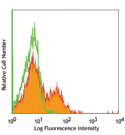
Compare Data Across All Formats
This data display is provided for general comparisons between formats.
Your actual data may vary due to variations in samples, target cells, instruments and their settings, staining conditions, and other factors.
If you need assistance with selecting the best format contact our expert technical support team.


Follow Us