 Login / Register


- Clone
- TUGh4 (See other available formats)
- Regulatory Status
- RUO
- Workshop
- VI C-89
- Other Names
- Common γ chain, γc, IL-2 receptor γ subunit
- Isotype
- Rat IgG2b, κ
- Ave. Rating
- Submit a Review
- Product Citations
- publications

-

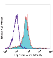
Human peripheral blood lymphocytes stained with TUGh4 APC
| Cat # | Size | Price | Quantity Check Availability | Save | ||
|---|---|---|---|---|---|---|
| 338607 | 25 tests | 114€ | ||||
| 338608 | 100 tests | 266€ | ||||
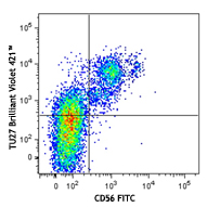
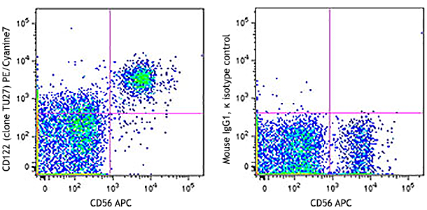
CD132 is a 64-70 kD type I transmembrane glycoprotein of the Ig superfamily, also known as common γ chain (γc), or IL-2 receptor γ subunit. It is expressed broadly on T- and B-lymphocytes, NK cells, monocytes, and granulocytes. CD132 is an essential component of cytokine receptors for IL-2, IL-4, IL-7, IL-9, IL-15 and IL-21. Ligand binding induces tyrosine phosphorylation and initiates signaling through a JAK/STAT pathway. CD132 mutation results in X-linked severe combined immune deficiency (XSCID).
Product DetailsProduct Details
- Reactivity
- Human
- Antibody Type
- Monoclonal
- Host Species
- Rat
- Formulation
- Phosphate-buffered solution, pH 7.2, containing 0.09% sodium azide and BSA (origin USA)
- Preparation
- The antibody was purified by affinity chromatography, and conjugated with APC under optimal conditions.
- Concentration
- Lot-specific (to obtain lot-specific concentration and expiration, please enter the lot number in our Certificate of Analysis online tool.)
- Storage & Handling
- The antibody solution should be stored undiluted between 2°C and 8°C, and protected from prolonged exposure to light. Do not freeze.
- Application
-
FC - Quality tested
- Recommended Usage
-
Each lot of this antibody is quality control tested by immunofluorescent staining with flow cytometric analysis. For flow cytometric staining, the suggested use of this reagent is 5 µl per million cells in 100 µl staining volume or 5 µl per 100 µl of whole blood.
- Excitation Laser
-
Red Laser (633 nm)
- Application References
-
- Itano M, et al. 1996. J. Exp. Med. 178:389
- Kondo M, et al. 1993. Science 262:1874
- Product Citations
- RRID
-
AB_2123585 (BioLegend Cat. No. 338607)
AB_2123584 (BioLegend Cat. No. 338608)
Antigen Details
- Structure
- Type I transmembrane glycoprotein, Ig superfamily 64-70 kD
- Distribution
-
T cells, B cells, NK, monocytes, granulocytes
- Ligand/Receptor
- Component of cytokine receptors for IL-2, IL-4, IL-7, IL-9, IL-15 and IL-21
- Cell Type
- B cells, Granulocytes, Monocytes, NK cells, T cells
- Biology Area
- Immunology
- Molecular Family
- CD Molecules, Cytokine/Chemokine Receptors
- Antigen References
-
1. Zola H, et al. eds. 2007. Leukocyte and Stromal Cell Molecules:The CD Markers. Wiely-Liss A John Wiley & Sons Inc, Publication
2. Nakarai T, et al. 1994. J. Exp. Med. 180:241
3. Kawahara A, et al. 1995. Proc. Natl. Acad. Sci. USA. 92:8724
4. Habib T, et al. 2002. Biochemistry. 41:8725
5. Matthews DJ, et al. 1995. Blood 85:38 - Gene ID
- 3561 View all products for this Gene ID
- UniProt
- View information about CD132 on UniProt.org
Related Pages & Pathways
Pathways


Customers Also Purchased

Compare Data Across All Formats
This data display is provided for general comparisons between formats.
Your actual data may vary due to variations in samples, target cells, instruments and their settings, staining conditions, and other factors.
If you need assistance with selecting the best format contact our expert technical support team.


Follow Us