 Login / Register


- Clone
- A019D5 (See other available formats)
- Regulatory Status
- RUO
- Other Names
- IL-7 receptor α chain, IL-7Rα
- Isotype
- Mouse IgG1, κ
- Ave. Rating
- Submit a Review
- Product Citations
- publications

-

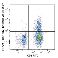
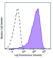
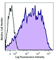
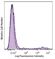
Human peripheral blood lymphocytes were stained with CD3 FITC and CD127 (clone A019D5) Brilliant Violet 605™ (top) or mouse IgG1, κ Brilliant Violet 605™ isotype control (bottom). -

| Cat # | Size | Price | Quantity Check Availability | Save | ||
|---|---|---|---|---|---|---|
| 351333 | 25 tests | 181€ | ||||
| 351334 | 100 tests | 357€ | ||||
CD127 is a 60-90 kD type I transmembrane glycoprotein also known as IL-7 receptor α chain or IL-7Rα. It forms a heterodimer with the common γ chain (γc or CD132) which is shared with the receptors for IL-2, IL-4, IL-9, IL-13, IL-15, and IL-21. CD127 is expressed on immature B cells through early pre-B stage cells, thymocytes (except CD4/CD8 double positive thymocytes), peripheral T cells, and bone marrow stromal cells. CD127 has been reported to be a useful marker for identifying memory and effector T cells. Studies have shown that CD127 expression is down-modulated on Treg cells. It can be used as a marker for differentiation of Treg and conventional T cells. The ligation of IL-7 with its receptor is important for stimulation of mature and immature T cells as well as immature B cell proliferation and development.
Product DetailsProduct Details
- Reactivity
- Human
- Antibody Type
- Monoclonal
- Host Species
- Mouse
- Immunogen
- Recombinant human CD127
- Formulation
- Phosphate-buffered solution, pH 7.2, containing 0.09% sodium azide and BSA (origin USA).
- Preparation
- The antibody was purified by affinity chromatography and conjugated with Brilliant Violet 605™ under optimal conditions.
- Concentration
- Lot-specific (to obtain lot-specific concentration and expiration, please enter the lot number in our Certificate of Analysis online tool.)
- Storage & Handling
- The antibody solution should be stored undiluted between 2°C and 8°C, and protected from prolonged exposure to light. Do not freeze.
- Application
-
FC - Quality tested
- Recommended Usage
-
Each lot of this antibody is quality control tested by immunofluorescent staining with flow cytometric analysis. For flow cytometric staining, the suggested use of this reagent is 5 µl per million cells in 100 µl staining volume or 5 µl per 100 µl of whole blood.
Brilliant Violet 605™ excites at 405 nm and emits at 603 nm. The bandpass filter 610/20 nm is recommended for detection, although filter optimization may be required depending on other fluorophores used. Be sure to verify that your cytometer configuration and software setup are appropriate for detecting this channel. Refer to your instrument manual or manufacturer for support. Brilliant Violet 605™ is a trademark of Sirigen Group Ltd.
Learn more about Brilliant Violet™.
This product is subject to proprietary rights of Sirigen Inc. and is made and sold under license from Sirigen Inc. The purchase of this product conveys to the buyer a non-transferable right to use the purchased product for research purposes only. This product may not be resold or incorporated in any manner into another product for resale. Any use for therapeutics or diagnostics is strictly prohibited. This product is covered by U.S. Patent(s), pending patent applications and foreign equivalents. - Excitation Laser
-
Violet Laser (405 nm)
- Application Notes
-
Additional reported (for the relevant formats) application: proteogenomics1.
- Application References
-
- Peterson VM, et al. 2017. Nat. Biotechnol. 35:936. (PG)
- Product Citations
- RRID
-
AB_2562019 (BioLegend Cat. No. 351333)
AB_2562022 (BioLegend Cat. No. 351334)
Antigen Details
- Structure
- Type I transmembrane glycoprotein, associates with CD132, 60-90 kD
- Distribution
-
Immature B cells through early pre-B stage, thymocytes (except CD4/CD8 double positive thymocytes), peripheral T cells, bone marrow stromal cells
- Function
- T cell and immature B cell proliferation and development
- Ligand/Receptor
- IL-7
- Cell Type
- B cells, T cells, Thymocytes, Tregs
- Biology Area
- Immunology
- Molecular Family
- CD Molecules, Cytokine/Chemokine Receptors
- Antigen References
-
1. Sudo T, et al. 1993. P. Natl. Acad. Sci. USA 90:9125.
2. He YW and Malek TR. 1998. Crit. Rev. Immunol. 18:503.
3. Huster KM, et al. 2004. P. Natl. Acad. Sci. USA 101:5610.
4. Pillai M, et al. 2004. Leukemia Lymphoma 45:2403.
5. Morrissey PJ, et al. 1989. J. Exp. Med. 169:707.
6. Liu W, et al. 2006. J. Exp. Med. 203:1701. - Gene ID
- 3575 View all products for this Gene ID
- UniProt
- View information about CD127 on UniProt.org
Related Pages & Pathways
Pathways


Customers Also Purchased
Compare Data Across All Formats
This data display is provided for general comparisons between formats.
Your actual data may vary due to variations in samples, target cells, instruments and their settings, staining conditions, and other factors.
If you need assistance with selecting the best format contact our expert technical support team.


Follow Us