 Login / Register


- Clone
- IM7 (See other available formats)
- Regulatory Status
- RUO
- Other Names
- Hermes, Pgp-1, H-CAM, HUTCH-1, ECMR III, gp85, Ly-24
- Isotype
- Rat IgG2b, κ
- Ave. Rating
- Submit a Review
- Product Citations
- publications

-

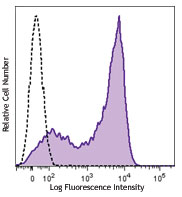
C57BL/6 mouse splenocytes were stained with CD44 (clone IM7) Brilliant Violet 785™.
| Cat # | Size | Price | Quantity Check Availability | Save | ||
|---|---|---|---|---|---|---|
| 103041 | 125 µL | 172€ | ||||
| 103059 | 50 µg | 219€ | ||||
CD44 is a 80-95 kD glycoprotein also known as Hermes, Pgp1, H-CAM, or HUTCH. It is expressed on all leukocytes, endothelial cells, hepatocytes, and mesenchymal cells. As B and T cells become activated or progress to the memory stage, CD44 expression increases from low or mid levels to high levels. Thus, CD44 has been reported to be a valuable marker for memory cell subsets. High CD44 expression on Treg cells has been associated with potent suppressive function via high production of IL-10. CD44 is an adhesion molecule involved in leukocyte attachment to and rolling on endothelial cells, homing to peripheral lymphoid organs and to the sites of inflammation, and leukocyte aggregation.
Product DetailsProduct Details
- Reactivity
- Mouse,Human
- Antibody Type
- Monoclonal
- Host Species
- Rat
- Immunogen
- Dexamethasone-induced myeloid leukemia M1 cells
- Formulation
- Phosphate-buffered solution, pH 7.2, containing 0.09% sodium azide and BSA (origin USA).
- Preparation
- The antibody was purified by affinity chromatography and conjugated with Brilliant Violet 785™ under optimal conditions.
- Concentration
- µg sizes: 0.2 mg/mLtest sizes: lot-specific (to obtain lot-specific concentration and expiration, please enter the lot number in our Certificate of Analysis online tool.)
- Storage & Handling
- The antibody solution should be stored undiluted between 2°C and 8°C, and protected from prolonged exposure to light. Do not freeze.
- Application
-
FC - Quality tested
- Recommended Usage
-
Each lot of this antibody is quality control tested by immunofluorescent staining with flow cytometric analysis. For flow cytometric staining using the test size, the suggested use of this reagent is 5 µl per million cells in 100 µl staining volume or 5 µl per 100 µl of whole blood. For flow cytometric staining using the µg size, the suggested use of this reagent is ≤0.4 µg per million cells in 100 µl volume. It is recommended that the reagent be titrated for optimal performance for each application.
Brilliant Violet 785™ excites at 405 nm and emits at 785 nm. The bandpass filter 780/60 nm is recommended for detection, although filter optimization may be required depending on other fluorophores used. Be sure to verify that your cytometer configuration and software setup are appropriate for detecting this channel. Refer to your instrument manual or manufacturer for support. Brilliant Violet 785™ is a trademark of Sirigen Group Ltd.
Learn more about Brilliant Violet™.
This product is subject to proprietary rights of Sirigen Inc. and is made and sold under license from Sirigen Inc. The purchase of this product conveys to the buyer a non-transferable right to use the purchased product for research purposes only. This product may not be resold or incorporated in any manner into another product for resale. Any use for therapeutics or diagnostics is strictly prohibited. This product is covered by U.S. Patent(s), pending patent applications and foreign equivalents. - Excitation Laser
-
Violet Laser (405 nm)
- Application Notes
-
Clone IM7 has been reported to recognize an epitope common to alloantigens and all isoforms of CD4417,18 that is located between amino acids 145 and 18620. This clone has been verified for immunocytochemistry (ICC) and frozen immunohistochemistry (IHC-F). Additional reported applications (for the relevant formats) include: immunohistochemistry of acetone-fixed frozen sections and formalin-fixed paraffin-embedded sections6,7, complement-mediated cytotoxicity1, immunoprecipitation1,3, in vivo inhibition of DTH4,5, and spatial biology (IBEX)23,24. The Ultra-LEAF™ purified antibody (Endotoxin < 0.01 EU/µg, Azide-Free, 0.2 µm filtered) is recommended for functional assays (Cat. No. 103046, 103065 - 103069).
Cross-reactivity to ferret has been reported by a collaborator, but not verified in house. - Application References
-
- Trowbridge IS, et al. 1982. Immunogenetics 15:299. (ICFC, IP, CMCD)
- Katoh S, et al. 1994. J. Immunol. 153:3440. (ELISA)
- Budd RC, et al. 1987. J. Immunol. 138:3120. (IP)
- Camp RL, et al. 1993. J. Exp. Med. 178:497. (Block)
- Weiss JM, et al. 1997. J. Cell Biol. 137:1137. (Block)
- Frank NY, et al. 2005. Cancer Res. 65:4320. (IHC) PubMed
- Cuff CA, et al. 2001. J. Clin. Invest. 108:1031. (IHC)
- Lee JW, et al. 2006. Nature Immunol. 8:181.
- Zhang N, et al. 2005. J. Immunol. 174:6967. PubMed
- Huabiao C, et al. 2005. J. Immunol. 175:591. PubMed
- Gui J, et al. 2007. Int. Immunol. 19:1201. PubMed
- Wang XY, et al. 2008. Blood 111:2436. PubMed
- Kenna TJ, et al. 2008. Blood 111:2091. PubMed
- Yamazaki J, et al. 2009. Blood PubMed
- Kmieciak M, et al. 2009. J. Transl. Med. 7:89. (FC) PubMed
- Chen YW, et al. 2010. Mol. Cancer Ther. 9:2879. PubMed
- Zheng Z, et al. 1995. J. Cell. Biol. 130:485.
- Wiranowska M, et al. 2010. Int. J. Cancer 127:532.
- Hirokawa Y, et al. 2014. Am J Physiol Gastrointerest Liver Physiol. 306:547. PubMed
- Sandmaier BM, et al. 1998. Blood 91:3494.
- Yang Y, et al. 2015. Hypertension. 65:1047. PubMed
- Peterson VM, et al. 2017. Nat. Biotechnol. 35:936. (PG)
- Radtke AJ, et al. 2020. Proc Natl Acad Sci U S A. 117:33455-65. (SB) PubMed
- Radtke AJ, et al. 2022. Nat Protoc. 17:378-401. (SB) PubMed
- Product Citations
- RRID
-
AB_11218802 (BioLegend Cat. No. 103041)
AB_2571953 (BioLegend Cat. No. 103059)
Antigen Details
- Structure
- Variable splicing of CD44 gene generates many CD44 isoforms, 80-95 kD
- Distribution
-
All leukocytes, epithelial cells, endothelial cells, hepatocytes, mesenchymal cells
- Function
- Leukocyte attachment and rolling on endothelial cells, stromal cells and ECM
- Ligand/Receptor
- Hyaluronan, MIP-1β, fibronectin, collagen
- Cell Type
- B cells, Endothelial cells, Epithelial cells, Leukocytes, Mesenchymal cells, Mesenchymal Stem Cells, Tregs
- Biology Area
- Cell Adhesion, Cell Biology, Immunology, Stem Cells
- Molecular Family
- Adhesion Molecules, CD Molecules
- Antigen References
-
1. Barclay AN, et al. 1997. The Leukocyte Antigen FactsBook Academic Press.
2. Haynes BF, et al. 1991. Cancer Cells 3:347.
3. Goldstein LA, et al. 1989. Cell 56:1063.
4. Mikecz K, et al. 1995. Nat. Med. 1:558.
5. Hegde V, et al. 2008. J. Leukocyte Biol. 84:134.
6. Liu T, et al. 2009. Biol. Direct 4:40. - Gene ID
- 12505 View all products for this Gene ID 960 View all products for this Gene ID
- UniProt
- View information about CD44 on UniProt.org
Related Pages & Pathways
Pathways


Customers Also Purchased
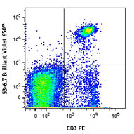
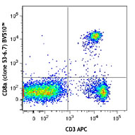
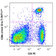
Compare Data Across All Formats
This data display is provided for general comparisons between formats.
Your actual data may vary due to variations in samples, target cells, instruments and their settings, staining conditions, and other factors.
If you need assistance with selecting the best format contact our expert technical support team.


Follow Us