 Login / Register


- Clone
- L3D10 (See other available formats)
- Regulatory Status
- RUO
- Other Names
- Cytotoxic T-Lymphocyte Antigen 4
- Isotype
- Mouse IgG1, κ
- Ave. Rating
- Submit a Review
- Product Citations
- publications

-

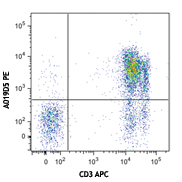
PMA/Ionomycin-stimulated human peripheral blood lymphocytes were stained with anti-human CD4 APC, then fixed, permeabilized and intracellularly stained with anti-CD152 (CTLA-4) (clone L3D10) PE (left) or Mouse IgG1, κ isotype control PE (right).
| Cat # | Size | Price | Quantity Check Availability | Save | ||
|---|---|---|---|---|---|---|
| 349905 | 25 tests | 94€ | ||||
| 349906 | 100 tests | 224€ | ||||
CD152, also known as Cytotoxic T-Lymphocyte Antigen 4 (CTLA-4), is a 33 kD member of the immunoglobulin superfamily. It is transiently expressed on activated T cells. CTLA4 is expressed on the surface of helper T cells and transmits an inhibitory signal to T cells. Regulatory T cells express high levels of CTLA-4. CTLA-4 (CD152) is similar to CD28 in amino acid sequence, structure, and genomic organization. Whereas CD28 delivers a costimulatory signal in T cell activation, CTLA-4 negatively regulates cell-mediated immune responses through interaction with CD80 (B7-1) and CD86 (B7-2) present on antigen presenting cells (APC). CTLA-4 is thought to play a role in the induction and maintenance of immunological tolerance as well as the development of protective immunity and thymocyte regulation.
Mutations in the CTLA-4 gene have been associated with various autoimmune diseases, such as systemic lupus erythematosus, insulin-dependent diabetes mellitus, and other autoimmune diseases. A transcript of the CTLA-4 gene that may represent a native soluble form of CTLA-4 (sCTLA-4) showed that eleven of twenty patients with autoimmune thyroid disease (ATD) had a high concentration of sCTLA-4, whereas only 1 of 30 apparently healthy volunteers contained measurable levels. sCTLA-4 immunoreactivity was inhibited by its binding to B7.1, suggesting that sCTLA-4 is a functional receptor. sCTLA4 also plays a role in the initial immune response to infection of immune cells by HIV, along with the CD-1 pathway and others.
Product Details
- Reactivity
- Human
- Antibody Type
- Monoclonal
- Host Species
- Mouse
- Immunogen
- Extracellular domain of human CTLA-4 and human IgG1 Fc fusion protein
- Formulation
- Phosphate-buffered solution, pH 7.2, containing 0.09% sodium azide and BSA (origin USA)
- Preparation
- The antibody was purified by affinity chromatography and conjugated with PE under optimal conditions.
- Concentration
- Lot-specific (to obtain lot-specific concentration and expiration, please enter the lot number in our Certificate of Analysis online tool.)
- Storage & Handling
- The antibody solution should be stored undiluted between 2°C and 8°C, and protected from prolonged exposure to light. Do not freeze.
- Application
-
ICFC - Quality tested
FC - Verified - Recommended Usage
-
Each lot of this antibody is quality control tested by intracellular immunofluorescent staining with flow cytometric analysis. For flow cytometric staining, the suggested use of this reagent is 5 µL per million cells in 100 µL staining volume or 5 µL per 100 µL of whole blood. It is recommended that the reagent be titrated for optimal performance for each application.
- Excitation Laser
-
Blue Laser (488 nm)
Green Laser (532 nm)/Yellow-Green Laser (561 nm)
- Application Notes
-
ELISA Detection: The biotinylated L3D10 antibody is useful as the detection antibody in a sandwich ELISA assay, when used in conjunction with the purified A3.6B10.G1 antibody (Cat. No. 525401) as the capture antibody and recombinant human CTLA-4 (Cat. No. 591909) as the standard.
Flow Cytometry: The fluorochrome-labeled L3D10 antibody is useful for immunofluorescent staining and flow cytometric analysis to identify CTLA-4-producing cells within mixed cell populations.
Note: For testing human soluble CTLA-4 in serum, plasma or cell culture supernatant, LEGEND MAX™ Human Soluble CTLA-4 ELISA Kit with Pre-coated Plates (Cat. No. 437407) are specially developed and recommended.
Additional reported applications (for the relevant formats) include: Blocking of CTLA-4/B7-1 interaction and blocking of CTLA-4-mediated inhibitory function to promote T cell expansion1,2.
This clone has been tested in-house and determined to not be suitable for applications in immunohistochemistry of frozen or paraffin-embedded tissue sections (IHC-F or IHC-P). - Application References
-
- May K, et al. 2005. Blood 105:1114. (Block)
- Lute K, et al. 2005. Blood 106:3127. (Block)
- Product Citations
- RRID
-
AB_10645522 (BioLegend Cat. No. 349905)
AB_10641842 (BioLegend Cat. No. 349906)
Antigen Details
- Structure
- Ig superfamily, 33 kD
- Distribution
-
Activated T cells and B cells
- Function
- Negative regulator of T cell activation
- Interaction
- Antigen presenting cells, such as dendritic cells
- Ligand/Receptor
- B7-1 (CD80), B7-2 (CD86)
- Cell Type
- B cells, T cells, Tregs
- Biology Area
- Immunology, Inhibitory Molecules
- Molecular Family
- CD Molecules, Immune Checkpoint Receptors
- Antigen References
-
1. Barclay N, et al. The Leukocyte Antigen FactsBook. Academic Press Inc. San Diego.
2. Kuiper H, et al. 1995. J. Immunol. 155:1776.
3. Lindsten T, et al. 1993. J. Immunol. 151:3489.
4. Morton P, et al. 1996. J. Immunol. 156:1047. - Gene ID
- 1493 View all products for this Gene ID
- UniProt
- View information about CD152 on UniProt.org
Related Pages & Pathways
Pathways


Related FAQs
- What type of PE do you use in your conjugates?
- We use R-PE in our conjugates.
Customers Also Purchased
Compare Data Across All Formats
This data display is provided for general comparisons between formats.
Your actual data may vary due to variations in samples, target cells, instruments and their settings, staining conditions, and other factors.
If you need assistance with selecting the best format contact our expert technical support team.


Follow Us