


- Clone
- EH12.2H7 (See other available formats)
- Regulatory Status
- RUO
- Other Names
- PD-1, PDCD1
- Isotype
- Mouse IgG1, κ
- Ave. Rating
- Submit a Review
- Product Citations
- publications

-

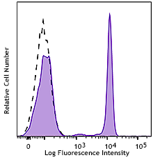
PHA-stimulated (day-3) human peripheral blood lymphocytes were stained with CD279 (clone EH12.2H7) PE/Cyanine7 (filled histogram) or mouse IgG1, κ PE/Cyanine7 (open histogram). -

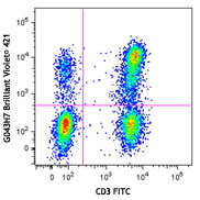
Human peripheral blood lymphocytes were stained with CD279 (clone EH12.2H7) PE/Cyanine7 and CD3 (clone UCHT1) FITC.
| Cat # | Size | Price | Save |
|---|---|---|---|
| 329917 | 25 tests | ¥38,720 | |
| 329918 | 100 tests | ¥84,040 |
Programmed cell death 1 (PD-1), also known as CD279, is a 55 kD member of the immunoglobulin superfamily. CD279 contains the immunoreceptor tyrosine-based inhibitory motif (ITIM) in the cytoplasmic region and plays a key role in peripheral tolerance and autoimmune disease. CD279 is expressed predominantly on activated T cells, B cells, and myeloid cells. PD-L1 (B7-H1) and PD-L2 (B7-DC) are ligands of CD279 (PD-1) and are members of the B7 gene family. Evidence suggests overlapping functions for these two PD-1 ligands and their constitutive expression on some normal tissues and upregulation on activated antigen-presenting cells. Interaction of CD279 ligands results in inhibition of T cell proliferation and cytokine secretion.
Product DetailsProduct Details
- Reactivity
- Human
- Antibody Type
- Monoclonal
- Host Species
- Mouse
- Formulation
- Phosphate-buffered solution, pH 7.2, containing 0.09% sodium azide and BSA (origin USA)
- Preparation
- The antibody was purified by affinity chromatography, and conjugated with PE/Cyanine7 under optimal conditions.
- Concentration
- Lot-specific (to obtain lot-specific concentration and expiration, please enter the lot number in our Certificate of Analysis online tool.)
- Storage & Handling
- The antibody solution should be stored undiluted between 2°C and 8°C, and protected from prolonged exposure to light. Do not freeze.
- Application
-
FC - Quality tested
- Recommended Usage
-
Each lot of this antibody is quality control tested by immunofluorescent staining with flow cytometric analysis. For flow cytometric staining, the suggested use of this reagent is 5 µl per million cells in 100 µl staining volume or 5 µl per 100 µl of whole blood.
- Excitation Laser
-
Blue Laser (488 nm)
Green Laser (532 nm)/Yellow-Green Laser (561 nm)
- Application Notes
-
Additional reported applications (for the relevant formats) include: blocking of ligand binding1-3, immunohistochemical staining of paraformaldehyde fixed frozen sections13, and spatial biology (IBEX)15,16. The LEAF™ purified antibody (Endotoxin <0.1 EU/µg, Azide-Free, 0.2 µm filtered) is recommended for functional assays (Cat. No. 329911 and 329912). For highly sensitive assays, we recommend Ultra-LEAF™ purified antibody (Cat. No. 329926) with a lower endotoxin limit than standard LEAF™ purified antibodies (Endotoxin <0.01 EU/µg).
- Additional Product Notes
- BioLegend is in the process of converting the name PE/Cy7 to PE/Cyanine7. The dye molecule remains the same, so you should expect the same quality and performance from our PE/Cyanine7 products. Please contact Technical Service if you have any questions.
-
Application References
(PubMed link indicates BioLegend citation) -
- Dorfman DM, et al. 2006 Am. J. Surg. Pathol. 30:802. (FA)
- Radziewicz H, et al. 2007. J. Virol. 81:2545. (FA)
- Velu V, et al. 2007. J. Virol. 81:5819. (FA)
- Zahn RC, et al. 2008. J. Virol. 82:11577. PubMed
- Chang WS, et al. 2008. J. Immunol. 181:6707. (FC) PubMed
- Nakamoto N, et al. 2009. PLoS Pathog. 5:e1000313. (FA)
- Jones RB, et al. 2009. J. Virol. 83:8722. (FC) PubMed
- Vojnov L, et al. 2010. J. Virol. 84:753. (FC) PubMed
- Radziewicz H, et al. 2010. J. Immunol. 184:2410. (FC) PubMed
- Monteriro P, et al. 2011. J. Immunol. 186:4618. PubMed
- Conrad J, et al. 2011. J. Immunol. 186:6871. PubMed
- Salisch NC, et al. 2010. J. Immunol. 184:476. (Rhesus reactivity)
- Li H and Pauza CD. 2015. Eur. J. Immunol. 45:298. (IHC)
- Peterson VM, et al. 2017. Nat. Biotechnol. 35:936. (PG)
- Radtke AJ, et al. 2020. Proc Natl Acad Sci USA. 117:33455-33465. (SB) PubMed
- Radtke AJ, et al. 2022. Nat Protoc. 17:378-401. (SB) PubMed
- Product Citations
- RRID
-
AB_2159325 (BioLegend Cat. No. 329917)
AB_2159324 (BioLegend Cat. No. 329918)
Antigen Details
- Structure
- Immunoglobulin superfamily
- Distribution
-
Transiently expressed on CD4- CD8- thymocytes; upregulated in thymocytes and splenic T and B lymphocytes; expressed on activated myeloid cells
- Ligand/Receptor
- B7-H1 (also known as PD-L1) and B7-DC (PD-L2)
- Cell Type
- B cells, Lymphocytes, T cells, Thymocytes, Tregs
- Biology Area
- Cancer Biomarkers, Immunology, Inhibitory Molecules
- Molecular Family
- CD Molecules, Immune Checkpoint Receptors
- Gene ID
- 5133 View all products for this Gene ID
- UniProt
- View information about CD279 on UniProt.org
Related Pages & Pathways
Pathways


Related FAQs
Customers Also Purchased
Compare Data Across All Formats
This data display is provided for general comparisons between formats.
Your actual data may vary due to variations in samples, target cells, instruments and their settings, staining conditions, and other factors.
If you need assistance with selecting the best format contact our expert technical support team.


Follow Us