


- Clone
- DREG-56 (See other available formats)
- Regulatory Status
- RUO
- Workshop
- V S056
- Other Names
- L-selectin, LECAM-1, LAM-1, Leu-8, TQ-1
- Isotype
- Mouse IgG1, κ
- Ave. Rating
- Submit a Review
- Product Citations
- publications

-

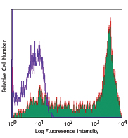
Human peripheral blood lymphocytes stained with DREG-56 PE/Cyanine7
| Cat # | Size | Price | Save |
|---|---|---|---|
| 304821 | 25 tests | ¥33,660 | |
| 304822 | 100 tests | ¥61,160 |
CD62L is a 74-95 kD single chain type I glycoprotein referred to as L-selectin or LECAM-1. It is expressed on most peripheral blood B cells, subsets of T and NK cells, monocytes, granulocytes, and certain hematopoietic malignant cells. CD62L binds to carbohydrates present on certain glycoforms of CD34, glycam-1, and MAdCAM-1 and with a low affinity to anionic oligosaccharide sequences related to sialylated Lewis X (sLex, CD15s) through its C-type lectin domain. CD62L is important for the homing of naïve lymphocytes to high endothelial venules in peripheral lymph nodes and Peyer's patches. It also plays a role in leukocyte rolling on activated endothelial cells.
Product DetailsProduct Details
- Reactivity
- Human
- Antibody Type
- Monoclonal
- Host Species
- Mouse
- Immunogen
- Concentrated supernatant from PMA-activated human peripheral blood leukocytes
- Formulation
- Phosphate-buffered solution, pH 7.2, containing 0.09% sodium azide and BSA (origin USA)
- Preparation
- The antibody was purified by affinity chromatography, and conjugated with PE/Cyanine7 under optimal conditions.
- Concentration
- Lot-specific (to obtain lot-specific concentration and expiration, please enter the lot number in our Certificate of Analysis online tool.)
- Storage & Handling
- The antibody solution should be stored undiluted between 2°C and 8°C, and protected from prolonged exposure to light. Do not freeze.
- Application
-
FC - Quality tested
- Recommended Usage
-
Each lot of this antibody is quality control tested by immunofluorescent staining with flow cytometric analysis. For flow cytometric staining, the suggested use of this reagent is 5 µl per million cells in 100 µl staining volume or 5 µl per 100 µl of whole blood.
- Excitation Laser
-
Blue Laser (488 nm)
Green Laser (532 nm)/Yellow-Green Laser (561 nm)
- Application Notes
-
Additional reported applications (for the relevant formats) include: Western blotting2,3,9 and in vitro blocking of lymphocytes binding to high endothelial venules (HEV)2. The Ultra-LEAF™ purified antibody (Endotoxin < 0.01 EU/µg, Azide-Free, 0.2 µm filtered) is recommended for functional assays (Cat. Nos. 304853-304858).
- Additional Product Notes
- BioLegend is in the process of converting the name PE/Cy7 to PE/Cyanine7. The dye molecule remains the same, so you should expect the same quality and performance from our PE/Cyanine7 products. Please contact Technical Service if you have any questions.
-
Application References
(PubMed link indicates BioLegend citation) -
- Schlossman S, et al. Eds. 1995. Leucocyte Typing V. Oxford University Press. New York.
- Kishimoto TK, et al. 1990. Proc. Natl. Acad. Sci. USA 87:2244. (WB, Block)
- Jutila M, et al. 2002. J. Immunol. 169:1768. (WB)
- Tamassia N, et al. 2008. J. Immunol. 181:6563. (FC) PubMed
- Kmieciak M, et al. 2009. J. Transl. Med. 7:89. (FC) PubMed
- Thakral D, et al. 2008. J. Immunol. 180:7431. (FC) PubMed
- Charles N, et al. 2010. Nat. Med. 16:701. (FC) PubMed
- Yoshino N, et al. 2000. Exp. Anim. (Tokyo) 49:97. (FC)
- Koenig JM, et al. 1996. Pediatr. Res. 39:616. (WB)
- Shi C, et al. 2011. J. Immunol. 187:5293. (FC) PubMed
- Burges M, et al. 2013. Clin Cancer Res. 19:5675. PubMed
- Cash JL, et al. 2013. EMBO Rep. 14:999. (FC) PubMed
- Product Citations
- RRID
-
AB_830800 (BioLegend Cat. No. 304821)
AB_830801 (BioLegend Cat. No. 304822)
Antigen Details
- Structure
- Selectin, single chain glycoprotein, 74-95 kD
- Distribution
-
Majority of B cells, naïve T cells, subset of memory T and NK cells, monocytes, granulocytes, thymocytes
- Function
- Leukocyte homing, leukocyte tethering, rolling
- Ligand/Receptor
- CD34, GlyCAM, MAdCAM-1
- Cell Type
- B cells, Granulocytes, Monocytes, Neutrophils, NK cells, T cells, Thymocytes, Tregs
- Biology Area
- Cell Adhesion, Cell Biology, Costimulatory Molecules, Immunology, Innate Immunity
- Molecular Family
- Adhesion Molecules, CD Molecules
- Antigen References
-
- Kishimoto T, et al. 1990. P. Natl. Acad. Sci. USA 87:2244.
- Kishimoto T, et al. 1991. Blood 78:805.
- Gene ID
- 6402 View all products for this Gene ID
- UniProt
- View information about CD62L on UniProt.org
Related Pages & Pathways
Pathways


Related FAQs
Customers Also Purchased
Compare Data Across All Formats
This data display is provided for general comparisons between formats.
Your actual data may vary due to variations in samples, target cells, instruments and their settings, staining conditions, and other factors.
If you need assistance with selecting the best format contact our expert technical support team.


Follow Us